1UA3 | pdb_00001ua3

Crystal structure of the pig pancreatic a-amylase complexed with malto-oligosaccharides


Experimental Data Snapshot

  • Method: X-RAY DIFFRACTION
  • Resolution: 2.01 Å
  • R-Value Free: 
    0.196 (Depositor) 
  • R-Value Work: 
    0.175 (Depositor), 0.162 (DCC) 
  • R-Value Observed: 
    0.183 (Depositor) 

wwPDB Validation 3D Report Full Report

Validation slider image for 1UA3

Ligand Structure Quality Assessment 


This is version 2.2 of the entry. See complete history

Literature

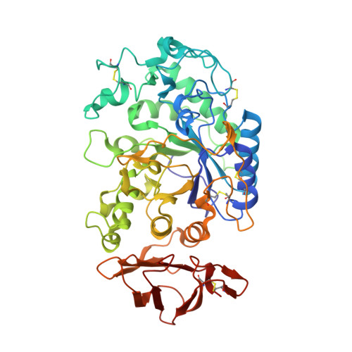
Crystal Structure of the Pig Pancreatic alpha-Amylase Complexed with Malto-Oligosaccharides

Payan, F.Qian, M.

(2003) J Protein Chem 22: 275-284

  • DOI: https://doi.org/10.1023/a:1025072520607
  • Primary Citation Related Structures: 
    1UA3

  • PubMed Abstract: 

    The structural X-ray map of a pig pancreatic alpha-amylase crystal soaked (and flash-frozen) with a maltopentaose substrate showed a pattern of electron density corresponding to the binding of oligosaccharides at the active site and at three surface binding sites. The electron density region observed at the active site, filling subsites-3 through-1, was interpreted in terms of the process of enzyme-catalyzed hydrolysis undergone by maltopentaose. Because the expected conformational changes in the "flexible loop" that constitutes the surface edge of the active site were not observed, the movement of the loop may depend on aglycone site being filled. The crystal structure was refined at 2.01 A resolution to an R factor of 17.0% ( R(free) factor of 19.8%). The final model consists of 3910 protein atoms, one calcium ion, two chloride ions, 103 oligosaccharide atoms, 761 atoms of water molecules, and 23 ethylene glycol atoms.


  • Organizational Affiliation
    • Architecture et Fonction des Macromolécules Biologiques, CNRS and Universities Aix-Marseille I and II, Marseille, France.

Macromolecule Content 

  • Total Structure Weight: 57.5 kDa 
  • Atom Count: 4,827 
  • Modeled Residue Count: 496 
  • Deposited Residue Count: 496 
  • Unique protein chains: 1

Macromolecules

Find similar proteins by:|  3D Structure
Entity ID: 1
MoleculeChains  Sequence LengthOrganismDetailsImage
Alpha-amylase, pancreatic496Sus scrofaMutation(s): 0 
EC: 3.2.1.1
UniProt
Find proteins for P00690 (Sus scrofa)
Explore P00690 
Go to UniProtKB:  P00690
Entity Groups
Sequence Clusters30% Identity50% Identity70% Identity90% Identity95% Identity100% Identity
UniProt GroupP00690
Sequence Annotations
Expand
Reference Sequence

Oligosaccharides

Help  
Entity ID: 2
MoleculeChains Length2D Diagram GlycosylationD Interactions
alpha-D-glucopyranose-(1-4)-alpha-D-glucopyranose
B
2N/A
Glycosylation Resources
GlyTouCan: G07411ON
GlyCosmos: G07411ON
Entity ID: 3
MoleculeChains Length2D Diagram GlycosylationD Interactions
alpha-D-glucopyranose-(1-4)-alpha-D-glucopyranose-(1-4)-alpha-D-glucopyranose
C, D
3N/A
Glycosylation Resources
GlyTouCan: G96370VA
GlyCosmos: G96370VA
GlyGen: G96370VA

Small Molecules

Ligands 4 Unique
IDChains Name / Formula / InChI Key2D Diagram3D Interactions
BGC

Query on BGC


Download:Ideal Coordinates CCD File
H [auth A]beta-D-glucopyranose
C6 H12 O6
WQZGKKKJIJFFOK-VFUOTHLCSA-N
EDO

Query on EDO


Download:Ideal Coordinates CCD File
I [auth A]
J [auth A]
K [auth A]
L [auth A]
M [auth A]
I [auth A],
J [auth A],
K [auth A],
L [auth A],
M [auth A],
N [auth A]
1,2-ETHANEDIOL
C2 H6 O2
LYCAIKOWRPUZTN-UHFFFAOYSA-N
CA

Query on CA


Download:Ideal Coordinates CCD File
G [auth A]CALCIUM ION
Ca
BHPQYMZQTOCNFJ-UHFFFAOYSA-N
CL

Query on CL


Download:Ideal Coordinates CCD File
E [auth A],
F [auth A]
CHLORIDE ION
Cl
VEXZGXHMUGYJMC-UHFFFAOYSA-M

Biologically Interesting Molecules (External Reference) 

2 Unique

Experimental Data & Validation

Experimental Data

  • Method: X-RAY DIFFRACTION
  • Resolution: 2.01 Å
  • R-Value Free:  0.196 (Depositor) 
  • R-Value Work:  0.175 (Depositor), 0.162 (DCC) 
  • R-Value Observed: 0.183 (Depositor) 
Space Group: P 21 21 21
Unit Cell:
Length ( Å )Angle ( ˚ )
a = 69.781α = 90
b = 113.183β = 90
c = 116.924γ = 90
Software Package:
Software NamePurpose
DENZOdata reduction
SCALEPACKdata scaling
AMoREphasing
CNSrefinement

Structure Validation

View Full Validation Report


Ligand Structure Quality Assessment 


Entry History 

Deposition Data

Revision History  (Full details and data files)

  • Version 1.0: 2003-10-14
    Type: Initial release
  • Version 1.1: 2008-04-27
    Changes: Version format compliance
  • Version 1.2: 2011-07-13
    Changes: Version format compliance
  • Version 2.0: 2020-07-29
    Type: Remediation
    Reason: Carbohydrate remediation
    Changes: Advisory, Atomic model, Data collection, Database references, Derived calculations, Non-polymer description, Structure summary
  • Version 2.1: 2023-12-27
    Changes: Data collection, Database references, Structure summary
  • Version 2.2: 2024-11-13
    Changes: Structure summary