1T60 | pdb_00001t60

Crystal structure of Type IV collagen NC1 domain from bovine lens capsule


Experimental Data Snapshot

  • Method: X-RAY DIFFRACTION
  • Resolution: 1.50 Å
  • R-Value Free: 
    0.216 (Depositor), 0.234 (DCC) 
  • R-Value Work: 
    0.189 (Depositor), 0.208 (DCC) 
  • R-Value Observed: 
    0.191 (Depositor) 

Starting Model: experimental
View more details

wwPDB Validation 3D Report Full Report

Validation slider image for 1T60

This is version 1.4 of the entry. See complete history

Literature

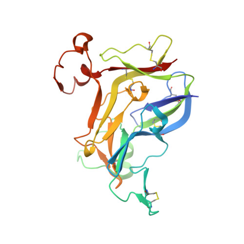
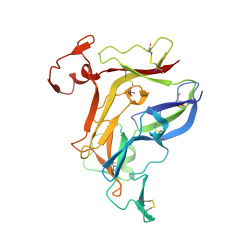
The alpha1.alpha2 network of collagen IV. Reinforced stabilization of the noncollagenous domain-1 by noncovalent forces and the absence of Met-Lys cross-links

Vanacore, R.M.Shanmugasundararaj, S.Friedman, D.B.Bondar, O.Hudson, B.G.Sundaramoorthy, M.

(2004) J Biological Chem 279: 44723-44730

  • DOI: https://doi.org/10.1074/jbc.M406344200
  • Primary Citation Related Structures: 
    1T60, 1T61

  • PubMed Abstract: 

    Collagen IV networks are present in all metazoa and underlie epithelia as a component of basement membranes. The networks are essential for tissue function and are defective in disease. They are assembled by the oligomerization of triple-helical protomers that are linked end-to-end. At the C terminus, two protomers are linked head-to-head by interactions of their trimeric noncollagenous domains, forming a hexamer structure. This linkage in the alpha1.alpha2 network is stabilized by a putative covalent Met-Lys cross-link between the trimer-trimer interface (Than, M. E., Henrich, S., Huber, R., Ries, A., Mann, K., Kuhn, K., Timpl, R., Bourenkov, G. P., Bartunik, H. D., and Bode, W. (2002) Proc. Natl. Acad. Sci. U. S. A. 99, 6607-6612) forming a nonreducible dimer that connects the hexamer. In the present study, this cross-link was further investigated by: (a) comparing the 1.5-A resolution crystal structures of the alpha1.alpha2 hexamers from bovine placenta and lens capsule basement membranes, (b) mass spectrometric analysis of monomer and nonreducible dimer subunits of placenta basement membrane hexamers, and (c) hexamer dissociation/re-association studies. The findings rule out the novel Met-Lys cross-link, as well as other covalent cross-links, but establish that the nonreducible dimer is an inherent structural feature of a subpopulation of hexamers. The dimers reflect the reinforced stabilization, by noncovalent forces, of the connection between two adjoining protomers of a network. The reinforcement extends to other types of collagen IV networks, and it underlies the cryptic nature of a B-cell epitope of the alpha3.alpha4.alpha5 hexamer, implicating the stabilization event in the etiology and pathogenesis of Goodpasture autoimmune disease.


  • Organizational Affiliation
    • Department of Biochemistry and Molecular Biology, Kansas University Medical Center, Kansas City, Kansas 66160, USA.

Macromolecule Content 

  • Total Structure Weight: 609.32 kDa 
  • Atom Count: 44,599 
  • Modeled Residue Count: 5,381 
  • Deposited Residue Count: 5,480 
  • Unique protein chains: 2

Macromolecules

Find similar proteins by:|  3D Structure
Entity ID: 1
MoleculeChains  Sequence LengthOrganismDetailsImage
type iv collagen
A, B, D, E, G
A, B, D, E, G, H, J, K, M, N, P, Q, S, T, V, W
229Bos taurusMutation(s): 0 
UniProt
Find proteins for Q7SIB2 (Bos taurus)
Explore Q7SIB2 
Go to UniProtKB:  Q7SIB2
Entity Groups
Sequence Clusters30% Identity50% Identity70% Identity90% Identity95% Identity100% Identity
UniProt GroupQ7SIB2
Sequence Annotations
Expand
Reference Sequence
Find similar proteins by:|  3D Structure
Entity ID: 2
MoleculeChains  Sequence LengthOrganismDetailsImage
type iv collagen
C, F, I, L, O
C, F, I, L, O, R, U, X
227Bos taurusMutation(s): 0 
UniProt
Find proteins for Q7SIB3 (Bos taurus)
Explore Q7SIB3 
Go to UniProtKB:  Q7SIB3
Entity Groups
Sequence Clusters30% Identity50% Identity70% Identity90% Identity95% Identity100% Identity
UniProt GroupQ7SIB3
Sequence Annotations
Expand
Reference Sequence

Small Molecules

Ligands 3 Unique
IDChains Name / Formula / InChI Key2D Diagram3D Interactions
MPD

Query on MPD


Download:Ideal Coordinates CCD File
AB [auth P]
DB [auth S]
EB [auth S]
GA [auth D]
GB [auth T]
AB [auth P],
DB [auth S],
EB [auth S],
GA [auth D],
GB [auth T],
OA [auth J],
TA [auth M],
UA [auth M],
Z [auth A]
(4S)-2-METHYL-2,4-PENTANEDIOL
C6 H14 O2
SVTBMSDMJJWYQN-YFKPBYRVSA-N
K

Query on K


Download:Ideal Coordinates CCD File
AA [auth B]
BA [auth C]
CA [auth C]
EA [auth D]
FB [auth T]
AA [auth B],
BA [auth C],
CA [auth C],
EA [auth D],
FB [auth T],
HB [auth U],
IA [auth G],
IB [auth U],
KA [auth H],
KB [auth V],
LA [auth I],
PA [auth L],
RA [auth M],
VA [auth N],
WA [auth O],
YA [auth P]
POTASSIUM ION
K
NPYPAHLBTDXSSS-UHFFFAOYSA-N
CL

Query on CL


Download:Ideal Coordinates CCD File
BB [auth R]
CB [auth S]
DA [auth C]
FA [auth D]
HA [auth F]
BB [auth R],
CB [auth S],
DA [auth C],
FA [auth D],
HA [auth F],
JA [auth G],
JB [auth U],
LB [auth V],
MA [auth I],
MB [auth X],
NA [auth J],
QA [auth L],
SA [auth M],
XA [auth O],
Y [auth A],
ZA [auth P]
CHLORIDE ION
Cl
VEXZGXHMUGYJMC-UHFFFAOYSA-M

Experimental Data & Validation

Experimental Data

  • Method: X-RAY DIFFRACTION
  • Resolution: 1.50 Å
  • R-Value Free:  0.216 (Depositor), 0.234 (DCC) 
  • R-Value Work:  0.189 (Depositor), 0.208 (DCC) 
  • R-Value Observed: 0.191 (Depositor) 
Space Group: P 1 21 1
Unit Cell:
Length ( Å )Angle ( ˚ )
a = 127.88α = 90
b = 140.129β = 91.26
c = 160.692γ = 90
Software Package:
Software NamePurpose
HKL-2000data collection
SCALEPACKdata scaling
AMoREphasing
CNSrefinement
HKL-2000data reduction

Structure Validation

View Full Validation Report


Entry History 

Deposition Data

Revision History  (Full details and data files)

  • Version 1.0: 2004-09-21
    Type: Initial release
  • Version 1.1: 2008-04-30
    Changes: Version format compliance
  • Version 1.2: 2011-07-13
    Changes: Version format compliance
  • Version 1.3: 2023-08-23
    Changes: Data collection, Database references, Derived calculations, Refinement description
  • Version 1.4: 2024-10-30
    Changes: Structure summary