1SA4 | pdb_00001sa4

human protein farnesyltransferase complexed with FPP and R115777


Experimental Data Snapshot

  • Method: X-RAY DIFFRACTION
  • Resolution: 2.10 Å
  • R-Value Free: 
    0.195 (Depositor), 0.189 (DCC) 
  • R-Value Work: 
    0.166 (Depositor), 0.174 (DCC) 
  • R-Value Observed: 
    0.166 (Depositor) 

Starting Model: experimental
View more details

wwPDB Validation 3D Report Full Report

Validation slider image for 1SA4

Ligand Structure Quality Assessment 


This is version 2.1 of the entry. See complete history

Literature

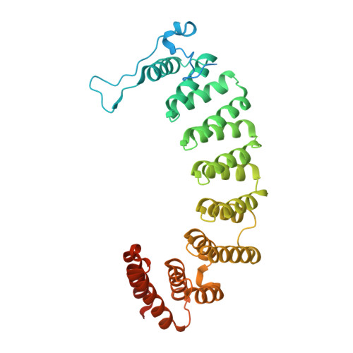
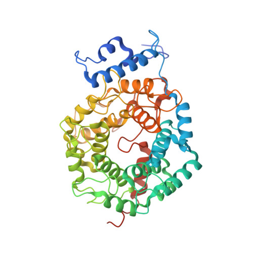
Crystal Structures of the Anticancer Clinical Candidates R115777 (Tipifarnib) and BMS-214662 Complexed with Protein Farnesyltransferase Suggest a Mechanism of FTI Selectivity.

Reid, T.S.Beese, L.S.

(2004) Biochemistry 43: 6877-6884

  • DOI: https://doi.org/10.1021/bi049723b
  • Primary Citation Related Structures: 
    1SA4, 1SA5

  • PubMed Abstract: 

    The search for new cancer therapeutics has identified protein farnesyltransferase (FTase) as a promising drug target. This enzyme attaches isoprenoid lipids to signal transduction proteins involved in growth and differentiation. The two FTase inhibitors (FTIs), R115777 (tipifarnib/Zarnestra) and BMS-214662, have undergone evaluation as cancer therapeutics in phase I and II clinical trials. R115777 has been evaluated in phase III clinical trials and shows indications for the treatment of blood and breast malignancies. Here we present crystal structures of R115777 and BMS-214662 complexed with mammalian FTase. These structures illustrate the molecular mechanism of inhibition and selectivity toward FTase over the related enzyme, protein geranylgeranyltransferase type I (GGTase-I). These results, combined with previous biochemical and structural analyses, identify features of FTase that could be exploited to modulate inhibitor potency and specificity and should aid in the continued development of FTIs as therapeutics for the treatment of cancer and parasitic infections.


  • Organizational Affiliation
    • Department of Biochemistry, Duke University Medical Center, Durham, North Carolina 27710, USA.

Macromolecule Content 

  • Total Structure Weight: 94.97 kDa 
  • Atom Count: 6,663 
  • Modeled Residue Count: 725 
  • Deposited Residue Count: 819 
  • Unique protein chains: 2

Macromolecules

Find similar proteins by:|  3D Structure
Entity ID: 1
MoleculeChains  Sequence LengthOrganismDetailsImage
Protein farnesyltransferase/geranylgeranyltransferase type I alpha subunit382Homo sapiensMutation(s): 0 
Gene Names: FNTA
EC: 2.5.1.58 (PDB Primary Data), 2.5.1.59 (PDB Primary Data)
UniProt & NIH Common Fund Data Resources
Find proteins for P49354 (Homo sapiens)
Explore P49354 
Go to UniProtKB:  P49354
PHAROS:  P49354
GTEx:  ENSG00000168522 
Entity Groups
Sequence Clusters30% Identity50% Identity70% Identity90% Identity95% Identity100% Identity
UniProt GroupP49354
Sequence Annotations
Expand
Reference Sequence
Find similar proteins by:|  3D Structure
Entity ID: 2
MoleculeChains  Sequence LengthOrganismDetailsImage
Protein farnesyltransferase beta subunit437Homo sapiensMutation(s): 0 
Gene Names: FNTB
EC: 2.5.1.58
UniProt & NIH Common Fund Data Resources
Find proteins for P49356 (Homo sapiens)
Explore P49356 
Go to UniProtKB:  P49356
PHAROS:  P49356
GTEx:  ENSG00000257365 
Entity Groups
Sequence Clusters30% Identity50% Identity70% Identity90% Identity95% Identity100% Identity
UniProt GroupP49356
Sequence Annotations
Expand
Reference Sequence

Oligosaccharides

Help  
Entity ID: 3
MoleculeChains Length2D Diagram GlycosylationD Interactions
beta-D-fructofuranose-(2-1)-alpha-D-glucopyranose
C
2N/A
Glycosylation Resources
GlyTouCan: G05551OP
GlyCosmos: G05551OP

Small Molecules

Ligands 3 Unique
IDChains Name / Formula / InChI Key2D Diagram3D Interactions
JAN

Query on JAN


Download:Ideal Coordinates CCD File
F [auth B]6-[(S)-AMINO(4-CHLOROPHENYL)(1-METHYL-1H-IMIDAZOL-5-YL)METHYL]-4-(3-CHLOROPHENYL)-1-METHYLQUINOLIN-2(1H)-ONE
C27 H22 Cl2 N4 O
PLHJCIYEEKOWNM-HHHXNRCGSA-N
FPP

Query on FPP


Download:Ideal Coordinates CCD File
E [auth B]FARNESYL DIPHOSPHATE
C15 H28 O7 P2
VWFJDQUYCIWHTN-YFVJMOTDSA-N
ZN

Query on ZN


Download:Ideal Coordinates CCD File
D [auth B]ZINC ION
Zn
PTFCDOFLOPIGGS-UHFFFAOYSA-N
Binding Affinity Annotations 
IDSourceBinding Affinity
JAN BindingDB:  1SA4 IC50: min: 0.65, max: 1100 (nM) from 4 assay(s)

Biologically Interesting Molecules (External Reference) 

1 Unique

Experimental Data & Validation

Experimental Data

  • Method: X-RAY DIFFRACTION
  • Resolution: 2.10 Å
  • R-Value Free:  0.195 (Depositor), 0.189 (DCC) 
  • R-Value Work:  0.166 (Depositor), 0.174 (DCC) 
  • R-Value Observed: 0.166 (Depositor) 
Space Group: P 61
Unit Cell:
Length ( Å )Angle ( ˚ )
a = 178.442α = 90
b = 178.442β = 90
c = 64.458γ = 120
Software Package:
Software NamePurpose
CNSrefinement
HKL-2000data reduction
HKL-2000data scaling
CNSphasing

Structure Validation

View Full Validation Report


Ligand Structure Quality Assessment 


Entry History 

Deposition Data

Revision History  (Full details and data files)

  • Version 1.0: 2004-06-08
    Type: Initial release
  • Version 1.1: 2008-04-29
    Changes: Version format compliance
  • Version 1.2: 2011-07-13
    Changes: Version format compliance
  • Version 2.0: 2020-07-29
    Type: Remediation
    Reason: Carbohydrate remediation
    Changes: Atomic model, Data collection, Database references, Derived calculations, Non-polymer description, Structure summary
  • Version 2.1: 2023-08-23
    Changes: Data collection, Database references, Refinement description, Structure summary