1R4I | pdb_00001r4i

Crystal Structure of Androgen Receptor DNA-Binding Domain Bound to a Direct Repeat Response Element


Experimental Data Snapshot

  • Method: X-RAY DIFFRACTION
  • Resolution: 3.10 Å
  • R-Value Free: 
    0.264 (Depositor), 0.262 (DCC) 
  • R-Value Work: 
    0.246 (Depositor), 0.243 (DCC) 

Starting Model: experimental
View more details

wwPDB Validation 3D Report Full Report

Validation slider image for 1R4I

This is version 1.5 of the entry. See complete history

Literature

Structural basis of androgen receptor binding to selective androgen response elements.

Shaffer, P.L.Jivan, A.Dollins, D.E.Claessens, F.Gewirth, D.T.

(2004) Proc Natl Acad Sci U S A 101: 4758-4763

  • DOI: https://doi.org/10.1073/pnas.0401123101
  • Primary Citation Related Structures: 
    1R4I

  • PubMed Abstract: 

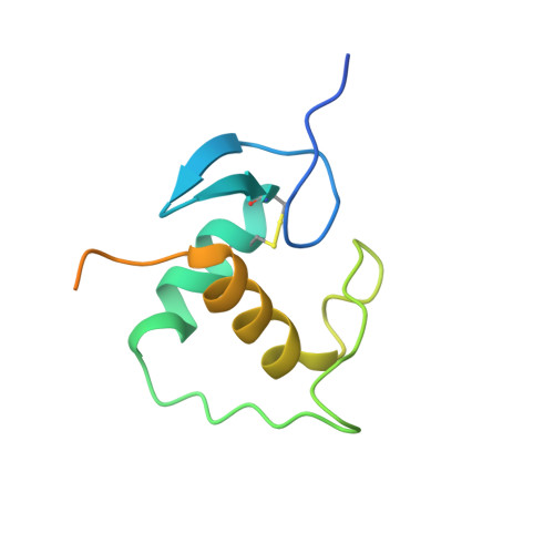
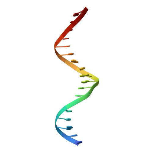
    Steroid receptors bind as dimers to a degenerate set of response elements containing inverted repeats of a hexameric half-site separated by 3 bp of spacer (IR3). Naturally occurring selective androgen response elements have recently been identified that resemble direct repeats of the hexameric half-site (ADR3). The 3D crystal structure of the androgen receptor (AR) DNA-binding domain bound to a selective ADR3 reveals an unexpected head-to-head arrangement of the two protomers rather than the expected head-to-tail arrangement seen in nuclear receptors bound to response elements of similar geometry. Compared with the glucocorticoid receptor, the DNA-binding domain dimer interface of the AR has additional interactions that stabilize the AR dimer and increase the affinity for nonconsensus response elements. This increased interfacial stability compared with the other steroid receptors may account for the selective binding of AR to ADR3 response elements.


  • Organizational Affiliation
    • Department of Biochemistry, Duke University Medical Center, Durham, NC 27710, USA.

Macromolecule Content 

  • Total Structure Weight: 34.59 kDa 
  • Atom Count: 1,813 
  • Modeled Residue Count: 181 
  • Deposited Residue Count: 246 
  • Unique protein chains: 1
  • Unique nucleic acid chains: 2

Macromolecules


Find similar proteins by:|  3D Structure
Entity ID: 3
MoleculeChains  Sequence LengthOrganismDetailsImage
Androgen receptorC [auth A],
D [auth B]
105Rattus norvegicusMutation(s): 1 
Gene Names: AR
UniProt
Find proteins for P15207 (Rattus norvegicus)
Explore P15207 
Go to UniProtKB:  P15207
Entity Groups
Sequence Clusters30% Identity50% Identity70% Identity90% Identity95% Identity100% Identity
UniProt GroupP15207
Sequence Annotations
Expand
Reference Sequence
Find similar nucleic acids by:  Sequence
Entity ID: 1
MoleculeChains LengthOrganismImage
5'-D(*CP*CP*AP*GP*AP*AP*CP*AP*TP*CP*AP*AP*GP*AP*AP*CP*AP*G)-3'A [auth C]18N/A
Sequence Annotations
Expand
Reference Sequence
Find similar nucleic acids by:  Sequence
Entity ID: 2
MoleculeChains LengthOrganismImage
5'-D(*CP*TP*GP*TP*TP*CP*TP*TP*GP*AP*TP*GP*TP*TP*CP*TP*GP*G)-3'B [auth D]18N/A
Sequence Annotations
Expand
Reference Sequence

Experimental Data & Validation

Experimental Data

  • Method: X-RAY DIFFRACTION
  • Resolution: 3.10 Å
  • R-Value Free:  0.264 (Depositor), 0.262 (DCC) 
  • R-Value Work:  0.246 (Depositor), 0.243 (DCC) 
Space Group: P 32 2 1
Unit Cell:
Length ( Å )Angle ( ˚ )
a = 137.89α = 90
b = 137.89β = 90
c = 85.71γ = 120
Software Package:
Software NamePurpose
CNSrefinement
HKL-2000data reduction
HKL-2000data scaling
SOLVEphasing
MOLREPphasing

Structure Validation

View Full Validation Report


Entry History 

Deposition Data

Revision History  (Full details and data files)

  • Version 1.0: 2004-06-29
    Type: Initial release
  • Version 1.1: 2008-04-29
    Changes: Version format compliance
  • Version 1.2: 2011-07-13
    Changes: Version format compliance
  • Version 1.3: 2021-10-27
    Changes: Database references, Derived calculations
  • Version 1.4: 2023-08-23
    Changes: Data collection, Refinement description
  • Version 1.5: 2024-10-16
    Changes: Structure summary