1R1A | pdb_00001r1a

CRYSTAL STRUCTURE OF HUMAN RHINOVIRUS SEROTYPE 1A (HRV1A)


Experimental Data Snapshot

  • Method: X-RAY DIFFRACTION
  • Resolution: 3.20 Å
  • R-Value Observed: 
    0.293 (Depositor) 

wwPDB Validation 3D Report Full Report

Validation slider image for 1R1A

This is version 3.2 of the entry. See complete history

Literature

Crystal structure of human rhinovirus serotype 1A (HRV1A).

Kim, S.S.Smith, T.J.Chapman, M.S.Rossmann, M.G.Pevear, D.C.Dutko, F.J.Felock, P.J.Diana, G.D.McKinlay, M.A.

(1989) J Mol Biology 210: 91-111

  • DOI: https://doi.org/10.1016/0022-2836(89)90293-3
  • Primary Citation Related Structures: 
    1R1A

  • PubMed Abstract: 

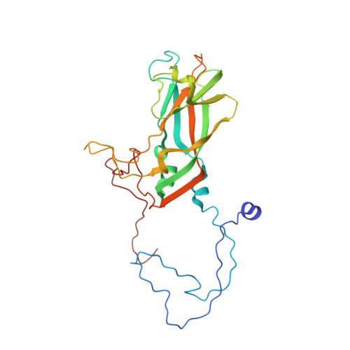
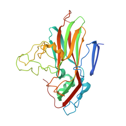
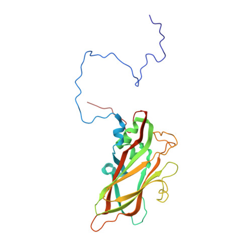
    The structure of human rhinovirus 1A (HRV1A) has been determined to 3.2 A resolution using phase refinement and extension by symmetry averaging starting with phases at 5 A resolution calculated from the known human rhinovirus 14 (HRV14) structure. The polypeptide backbone structures of HRV1A and HRV14 are similar, but the exposed surfaces are rather different. Differential charge distribution of amino acid residues in the "canyon", the putative receptor binding site, provides a possible explanation for the difference in minor versus major receptor group specificities, represented by HRV1A and HRV14, respectively. The hydrophobic pocket in VP1, into which antiviral compounds bind, is in an "open" conformation similar to that observed in drug-bound HRV14. Drug binding in HRV1A does not induce extensive conformational changes, in contrast to the case of HRV14.


  • Organizational Affiliation
    • Department of Biological Sciences, Purdue University, West Lafayette, IN 47907.

Macromolecule Content 

  • Total Structure Weight: 92.83 kDa 
  • Atom Count: 6,246 
  • Modeled Residue Count: 793 
  • Deposited Residue Count: 832 
  • Unique protein chains: 4

Macromolecules

Find similar proteins by:|  3D Structure
Entity ID: 1
MoleculeChains  Sequence LengthOrganismDetailsImage
HUMAN RHINOVIRUS 1A COAT PROTEIN (SUBUNIT VP1)A [auth 1]287Human rhinovirus 1AMutation(s): 0 
UniProt
Find proteins for P23008 (Human rhinovirus 1A)
Explore P23008 
Go to UniProtKB:  P23008
Entity Groups
Sequence Clusters30% Identity50% Identity70% Identity90% Identity95% Identity100% Identity
UniProt GroupP23008
Sequence Annotations
Expand
Reference Sequence
Find similar proteins by:|  3D Structure
Entity ID: 2
MoleculeChains  Sequence LengthOrganismDetailsImage
HUMAN RHINOVIRUS 1A COAT PROTEIN (SUBUNIT VP2)B [auth 2]263Human rhinovirus 1AMutation(s): 0 
UniProt
Find proteins for P23008 (Human rhinovirus 1A)
Explore P23008 
Go to UniProtKB:  P23008
Entity Groups
Sequence Clusters30% Identity50% Identity70% Identity90% Identity95% Identity100% Identity
UniProt GroupP23008
Sequence Annotations
Expand
Reference Sequence
Find similar proteins by:|  3D Structure
Entity ID: 3
MoleculeChains  Sequence LengthOrganismDetailsImage
HUMAN RHINOVIRUS 1A COAT PROTEIN (SUBUNIT VP3)C [auth 3]238Human rhinovirus 1AMutation(s): 0 
UniProt
Find proteins for P23008 (Human rhinovirus 1A)
Explore P23008 
Go to UniProtKB:  P23008
Entity Groups
Sequence Clusters30% Identity50% Identity70% Identity90% Identity95% Identity100% Identity
UniProt GroupP23008
Sequence Annotations
Expand
Reference Sequence
Find similar proteins by:|  3D Structure
Entity ID: 4
MoleculeChains  Sequence LengthOrganismDetailsImage
HUMAN RHINOVIRUS 1A COAT PROTEIN (SUBUNIT VP4)D [auth 4]44Human rhinovirus 1AMutation(s): 0 
UniProt
Find proteins for P23008 (Human rhinovirus 1A)
Explore P23008 
Go to UniProtKB:  P23008
Entity Groups
Sequence Clusters30% Identity50% Identity70% Identity90% Identity95% Identity100% Identity
UniProt GroupP23008
Sequence Annotations
Expand
Reference Sequence

Oligosaccharides

Help  
Entity ID: 5
MoleculeChains Length2D Diagram GlycosylationD Interactions
beta-D-fructofuranose-(2-1)-alpha-D-glucopyranoseE [auth A]2N/A
Glycosylation Resources
GlyTouCan: G05551OP
GlyCosmos: G05551OP

Biologically Interesting Molecules (External Reference) 

1 Unique
Entity ID: 5
IDChains NameType/Class2D Diagram3D Interactions
PRD_900003
Query on PRD_900003
E [auth A]sucroseOligosaccharide / Nutrient

Experimental Data & Validation

Experimental Data

  • Method: X-RAY DIFFRACTION
  • Resolution: 3.20 Å
  • R-Value Observed: 0.293 (Depositor) 
Space Group: P 63 2 2
Unit Cell:
Length ( Å )Angle ( ˚ )
a = 341.3α = 90
b = 341.3β = 90
c = 465.9γ = 120
Software Package:
Software NamePurpose
PROLSQrefinement

Structure Validation

View Full Validation Report


Entry History 

Deposition Data

Revision History  (Full details and data files)

  • Version 1.0: 1990-07-15
    Type: Initial release
  • Version 1.1: 2008-03-24
    Changes: Version format compliance
  • Version 1.2: 2011-07-13
    Changes: Version format compliance
  • Version 1.3: 2011-12-07
    Changes: Database references
  • Version 2.0: 2020-07-29
    Type: Remediation
    Reason: Carbohydrate remediation
    Changes: Advisory, Atomic model, Data collection, Derived calculations, Non-polymer description, Other, Structure summary
  • Version 3.0: 2023-02-08
    Type: Remediation
    Changes: Advisory, Atomic model, Data collection, Database references, Derived calculations, Other, Refinement description, Structure summary
  • Version 3.1: 2023-03-15
    Changes: Advisory
  • Version 3.2: 2024-12-25
    Changes: Advisory, Data collection, Derived calculations, Structure summary