1QKZ | pdb_00001qkz

Fab fragment (MN14C11.6) in complex with a peptide antigen derived from Neisseria meningitidis P1.7 serosubtype antigen and domain II from Streptococcal protein G


Experimental Data Snapshot

  • Method: X-RAY DIFFRACTION
  • Resolution: 1.95 Å
  • R-Value Free: 
    0.262 (Depositor) 
  • R-Value Work: 
    0.205 (Depositor) 
  • R-Value Observed: 
    0.209 (Depositor) 

Starting Model: experimental
View more details

wwPDB Validation 3D Report Full Report

Validation slider image for 1QKZ

This is version 1.4 of the entry. See complete history

Literature

Crystal Structure of an Fab Fragment in Complex with a Meningococcal Serosubtype Antigen and a Protein G Domain

Derrick, J.P.Feavers, I.M.Maiden, M.C.J.

(1999) J Mol Biology 293: 81

  • DOI: https://doi.org/10.1006/jmbi.1999.3144
  • Primary Citation Related Structures: 
    1QKZ

  • PubMed Abstract: 

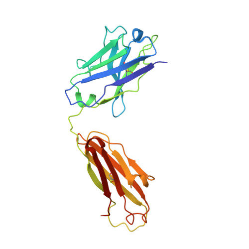
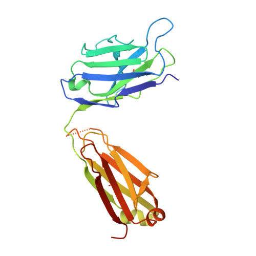
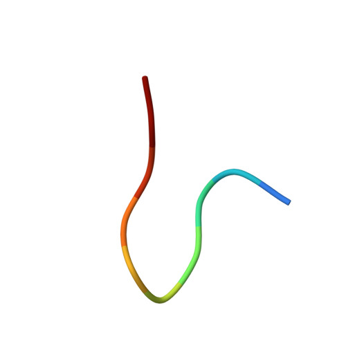
    Many pathogens present highly variable surface proteins to their host as a means of evading immune responses. The structure of a peptide antigen corresponding to the subtype P1.7 variant of the porin PorA from the human pathogen Neisseria meningitidis was determined by solution of the X-ray crystal structure of the ternary complex of the peptide (ANGGASGQVK) in complex with a Fab fragment and a domain from streptococcal protein G to 1.95 A resolution. The peptide adopted a beta-hairpin structure with a type I beta-turn between residues Gly4P and Gly7P, the conformation of the peptide being further stabilised by a pair of hydrogen bonds from the side-chain of Asn2P to main-chain atoms in Val9P. The antigen binding site within the Fab formed a distinct crevice lined by a high proportion of apolar amino acids. Recognition was supplemented by hydrogen bonds from heavy chain residues Thr50H, Asp95H, Leu97H and Tyr100H to main-chain and side-chain atoms in the peptide. Complementarity-determining region (CDR) 3 of the heavy chain was responsible for approximately 50 % of the buried surface area formed by peptide-Fab binding, with the remainder made up from CDRs 1 and 3 of the light chain and CDRs 1 and 2 of the heavy chain. Knowledge of the structures of variable surface antigens such as PorA is an essential prerequisite to a molecular understanding of antigenic variation and its implications for vaccine design.


  • Organizational Affiliation
    • Department of Biomolecular Sciences, UMIST, Manchester, M60 1QD, UK. Jeremy.Derrick@umist.ac.uk

Macromolecule Content 

  • Total Structure Weight: 55.61 kDa 
  • Atom Count: 4,369 
  • Modeled Residue Count: 501 
  • Deposited Residue Count: 510 
  • Unique protein chains: 4

Macromolecules

Find similar proteins by:|  3D Structure
Entity ID: 1
MoleculeChains  Sequence LengthOrganismDetailsImage
PROTEIN G-PRIME64Streptococcus sp. 'group GMutation(s): 1 
UniProt
Find proteins for P19909 (Streptococcus sp. group G)
Explore P19909 
Go to UniProtKB:  P19909
Entity Groups
Sequence Clusters30% Identity50% Identity70% Identity90% Identity95% Identity100% Identity
UniProt GroupP19909
Sequence Annotations
Expand
Reference Sequence
Find similar proteins by:|  3D Structure
Entity ID: 2
MoleculeChains  Sequence LengthOrganismDetailsImage
ANTIBODYB [auth H]219Mus musculusMutation(s): 0 
Find similar proteins by:|  3D Structure
Entity ID: 3
MoleculeChains  Sequence LengthOrganismDetailsImage
ANTIBODYC [auth L]217Mus musculusMutation(s): 0 
Find similar proteins by:  Sequence   |   3D Structure  
Entity ID: 4
MoleculeChains  Sequence LengthOrganismDetailsImage
MAJOR OUTER MEMBRANE PROTEIN P1.16D [auth P]10Neisseria meningitidisMutation(s): 0 
UniProt
Find proteins for P57041 (Neisseria meningitidis serogroup A / serotype 4A (strain DSM 15465 / Z2491))
Explore P57041 
Go to UniProtKB:  P57041
Entity Groups
Sequence Clusters30% Identity50% Identity70% Identity90% Identity95% Identity100% Identity
UniProt GroupP57041
Sequence Annotations
Expand
Reference Sequence

Experimental Data & Validation

Experimental Data

  • Method: X-RAY DIFFRACTION
  • Resolution: 1.95 Å
  • R-Value Free:  0.262 (Depositor) 
  • R-Value Work:  0.205 (Depositor) 
  • R-Value Observed: 0.209 (Depositor) 
Space Group: P 1 21 1
Unit Cell:
Length ( Å )Angle ( ˚ )
a = 43.482α = 90
b = 63.099β = 81.54
c = 89.374γ = 90
Software Package:
Software NamePurpose
REFMACrefinement
DENZOdata reduction
SCALEPACKdata scaling
AMoREphasing

Structure Validation

View Full Validation Report


Entry History 

Deposition Data

Revision History  (Full details and data files)

  • Version 1.0: 2000-02-06
    Type: Initial release
  • Version 1.1: 2011-05-08
    Changes: Version format compliance
  • Version 1.2: 2011-07-13
    Changes: Version format compliance
  • Version 1.3: 2023-12-13
    Changes: Data collection, Database references, Refinement description
  • Version 1.4: 2024-11-20
    Changes: Structure summary