1QHV | pdb_00001qhv

HUMAN ADENOVIRUS SEROTYPE 2 FIBRE HEAD


Experimental Data Snapshot

  • Method: X-RAY DIFFRACTION
  • Resolution: 1.51 Å
  • R-Value Free: 
    0.143 (Depositor), 0.168 (DCC) 
  • R-Value Work: 
    0.109 (Depositor) 

Starting Model: experimental
View more details

wwPDB Validation 3D Report Full Report

Validation slider image for 1QHV

This is version 1.3 of the entry. See complete history

Literature

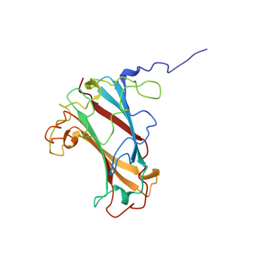
Structure of the human adenovirus serotype 2 fiber head domain at 1.5 A resolution.

van Raaij, M.J.Louis, N.Chroboczek, J.Cusack, S.

(1999) Virology 262: 333-343

  • DOI: https://doi.org/10.1006/viro.1999.9849
  • Primary Citation Related Structures: 
    1QHV

  • PubMed Abstract: 

    Adenovirus binds to its receptor via the head domain of its fiber protein. We have crystallized the adenovirus serotype 2 (subgroup C) receptor binding domain and solved the structure at 1.5 A resolution by the molecular replacement technique using the known adenovirus type 5 head structure. Included in the high-resolution model are 306 water molecules, five alternative side chain conformations, and individual anisotropic temperature factors for each atom. The overall structure of the serotype 2 head is very similar to its serotype 5 homologue, apart from differences in some of the flexible loops. All but subgroup B adenoviruses are believed to use the recently identified protein CAR (Coxsackievirus and adenovirus receptor) as receptor. By comparison of the two structures and sequence alignment of CAR binding and non-CAR binding serotype fiber heads, we discuss possible receptor binding sites and propose a receptor binding site in a crevice between two monomers on the side of the trimer. The structural basis of the extraordinary stability of the fiber head trimer is also discussed.


  • Organizational Affiliation
    • c/o Institut Laue Langevin, 6 rue Jules Horowitz, Grenoble Cedex 9, 38000, France.

Macromolecule Content 

  • Total Structure Weight: 21.67 kDa 
  • Atom Count: 1,836 
  • Modeled Residue Count: 195 
  • Deposited Residue Count: 195 
  • Unique protein chains: 1

Macromolecules

Find similar proteins by:|  3D Structure
Entity ID: 1
MoleculeChains  Sequence LengthOrganismDetailsImage
PROTEIN (ADENOVIRUS FIBRE)195Human adenovirus 2Mutation(s): 0 
Gene Names: LOCUS AD2H2
UniProt
Find proteins for P03275 (Human adenovirus C serotype 2)
Explore P03275 
Go to UniProtKB:  P03275
Entity Groups
Sequence Clusters30% Identity50% Identity70% Identity90% Identity95% Identity100% Identity
UniProt GroupP03275
Sequence Annotations
Expand
Reference Sequence

Experimental Data & Validation

Experimental Data

  • Method: X-RAY DIFFRACTION
  • Resolution: 1.51 Å
  • R-Value Free:  0.143 (Depositor), 0.168 (DCC) 
  • R-Value Work:  0.109 (Depositor) 
Space Group: P 3 2 1
Unit Cell:
Length ( Å )Angle ( ˚ )
a = 95.35α = 90
b = 95.35β = 90
c = 48.8γ = 120
Software Package:
Software NamePurpose
AMoREphasing
REFMACrefinement
MOSFLMdata reduction
CCP4data scaling

Structure Validation

View Full Validation Report


Entry History 

Deposition Data

Revision History  (Full details and data files)

  • Version 1.0: 1999-09-29
    Type: Initial release
  • Version 1.1: 2008-04-26
    Changes: Version format compliance
  • Version 1.2: 2011-07-13
    Changes: Derived calculations, Version format compliance
  • Version 1.3: 2023-08-16
    Changes: Data collection, Database references, Derived calculations, Refinement description