1QG0 | pdb_00001qg0

WILD-TYPE PEA FNR


Experimental Data Snapshot

  • Method: X-RAY DIFFRACTION
  • Resolution: 2.50 Å
  • R-Value Free: 
    0.273 (Depositor) 
  • R-Value Work: 
    0.218 (Depositor), 0.222 (DCC) 

Starting Model: experimental
View more details

wwPDB Validation 3D Report Full Report

Validation slider image for 1QG0

Ligand Structure Quality Assessment 


This is version 1.3 of the entry. See complete history

Literature

A productive NADP+ binding mode of ferredoxin-NADP+ reductase revealed by protein engineering and crystallographic studies.

Deng, Z.Aliverti, A.Zanetti, G.Arakaki, A.K.Ottado, J.Orellano, E.G.Calcaterra, N.B.Ceccarelli, E.A.Carrillo, N.Karplus, P.A.

(1999) Nat Struct Biol 6: 847-853

  • DOI: https://doi.org/10.1038/12307
  • Primary Citation Related Structures: 
    1QFY, 1QFZ, 1QG0, 1QGA

  • PubMed Abstract: 

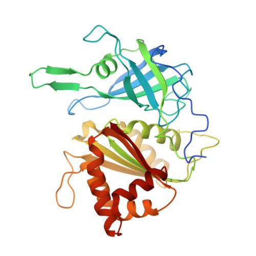
    The flavoenzyme ferredoxin-NADP+ reductase (FNR) catalyzes the production of NADPH during photosynthesis. Whereas the structures of FNRs from spinach leaf and a cyanobacterium as well as many of their homologs have been solved, none of these studies has yielded a productive geometry of the flavin-nicotinamide interaction. Here, we show that this failure occurs because nicotinamide binding to wild type FNR involves the energetically unfavorable displacement of the C-terminal Tyr side chain. We used mutants of this residue (Tyr 308) of pea FNR to obtain the structures of productive NADP+ and NADPH complexes. These structures reveal a unique NADP+ binding mode in which the nicotinamide ring is not parallel to the flavin isoalloxazine ring, but lies against it at an angle of approximately 30 degrees, with the C4 atom 3 A from the flavin N5 atom.


  • Organizational Affiliation
    • Section of Biochemistry, Molecular and Cell Biology, Cornell University, Ithaca, New York 14853, USA.

Macromolecule Content 

  • Total Structure Weight: 71.26 kDa 
  • Atom Count: 5,030 
  • Modeled Residue Count: 592 
  • Deposited Residue Count: 616 
  • Unique protein chains: 1

Macromolecules

Find similar proteins by:|  3D Structure
Entity ID: 1
MoleculeChains  Sequence LengthOrganismDetailsImage
PROTEIN (FERREDOXIN:NADP+ REDUCTASE)
A, B
308Lathyrus oleraceusMutation(s): 0 
EC: 1.18.1.2
UniProt
Find proteins for P10933 (Pisum sativum)
Explore P10933 
Go to UniProtKB:  P10933
Entity Groups
Sequence Clusters30% Identity50% Identity70% Identity90% Identity95% Identity100% Identity
UniProt GroupP10933
Sequence Annotations
Expand
Reference Sequence

Experimental Data & Validation

Experimental Data

  • Method: X-RAY DIFFRACTION
  • Resolution: 2.50 Å
  • R-Value Free:  0.273 (Depositor) 
  • R-Value Work:  0.218 (Depositor), 0.222 (DCC) 
Space Group: P 21 21 21
Unit Cell:
Length ( Å )Angle ( ˚ )
a = 73.9α = 90
b = 94.1β = 90
c = 140.2γ = 90
Software Package:
Software NamePurpose
DENZOdata reduction
SCALEPACKdata scaling
AMoREphasing
X-PLORrefinement

Structure Validation

View Full Validation Report


Ligand Structure Quality Assessment 


Entry History 

Deposition Data

Revision History  (Full details and data files)

  • Version 1.0: 1999-04-27
    Type: Initial release
  • Version 1.1: 2008-04-26
    Changes: Version format compliance
  • Version 1.2: 2011-07-13
    Changes: Version format compliance
  • Version 1.3: 2023-08-16
    Changes: Data collection, Database references, Derived calculations, Refinement description