1QFZ | pdb_00001qfz

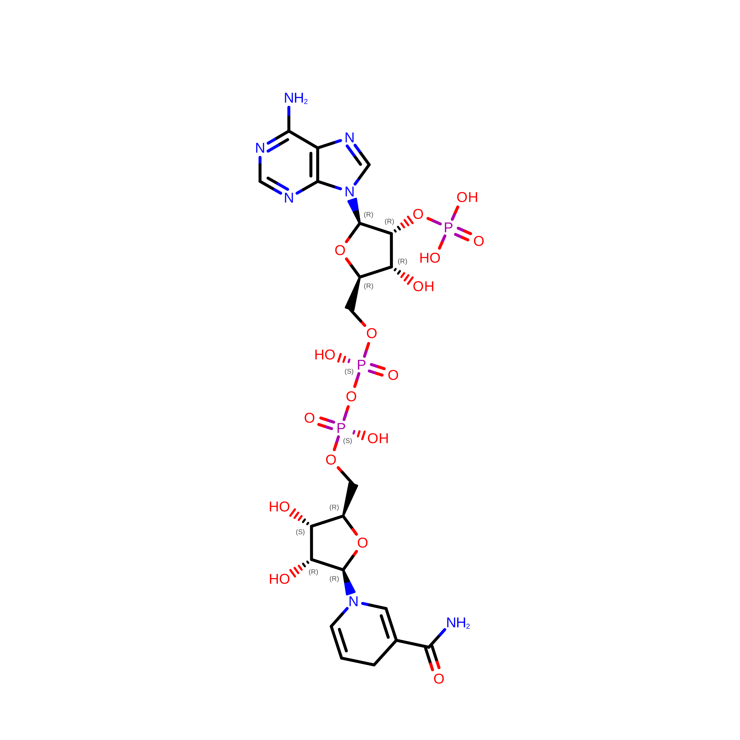
PEA FNR Y308S MUTANT IN COMPLEX WITH NADPH


Experimental Data Snapshot

  • Method: X-RAY DIFFRACTION
  • Resolution: 1.70 Å
  • R-Value Free: 
    0.235 (Depositor) 
  • R-Value Work: 
    0.201 (Depositor), 0.209 (DCC) 

Starting Model: experimental
View more details

wwPDB Validation 3D Report Full Report

Validation slider image for 1QFZ

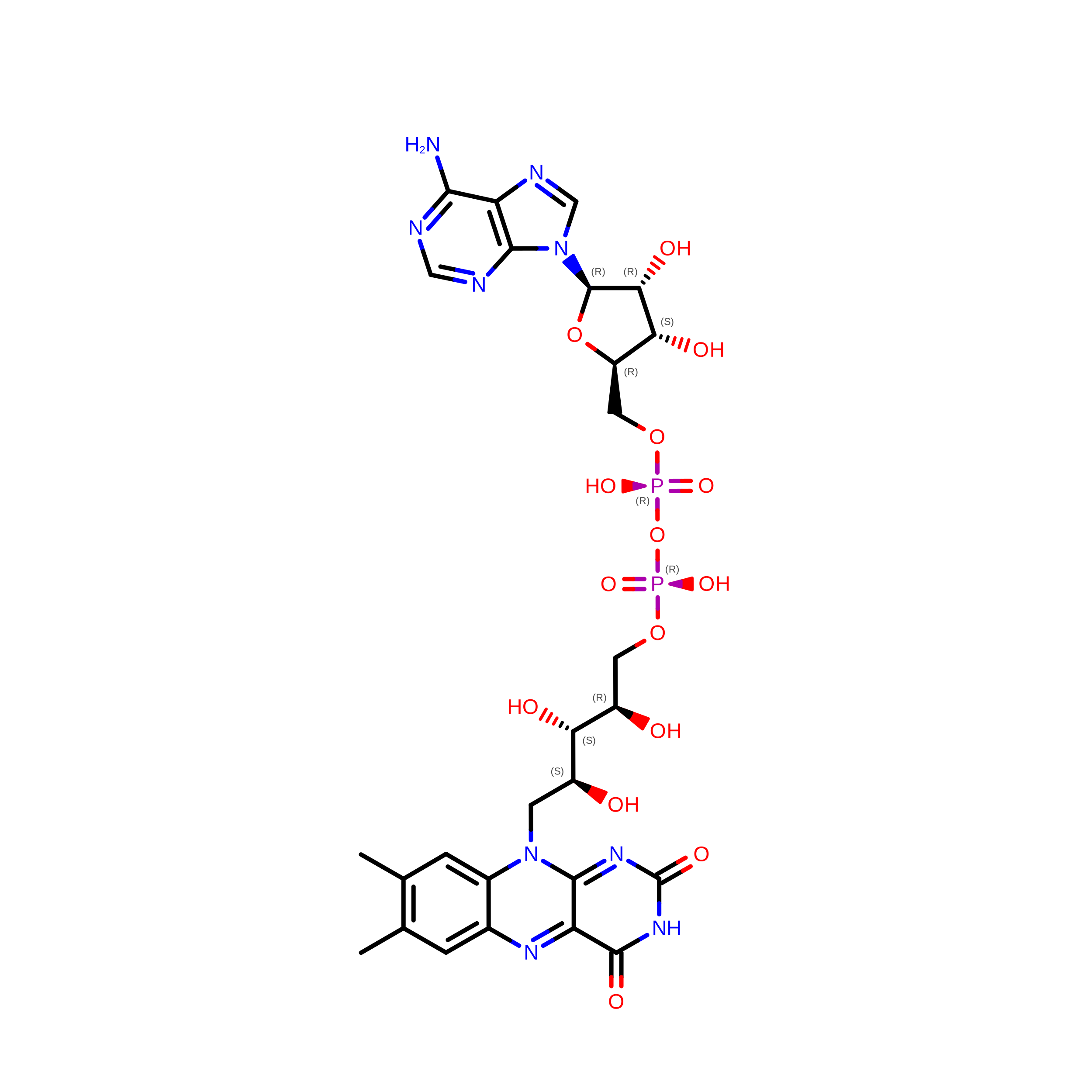
Ligand Structure Quality Assessment 


This is version 1.5 of the entry. See complete history. 

Literature

A productive NADP+ binding mode of ferredoxin-NADP+ reductase revealed by protein engineering and crystallographic studies.

Deng, Z., Aliverti, A., Zanetti, G., Arakaki, A.K., Ottado, J., Orellano, E.G., Calcaterra, N.B., Ceccarelli, E.A., Carrillo, N., Karplus, P.A.

(1999) Nat Struct Biol 6: 847-853

  • DOI: https://doi.org/10.1038/12307
  • Primary Citation Related Structures: 
    1QFY, 1QFZ, 1QG0, 1QGA

  • PubMed Abstract: 

    The flavoenzyme ferredoxin-NADP+ reductase (FNR) catalyzes the production of NADPH during photosynthesis. Whereas the structures of FNRs from spinach leaf and a cyanobacterium as well as many of their homologs have been solved, none of these studies has yielded a productive geometry of the flavin-nicotinamide interaction. Here, we show that this failure occurs because nicotinamide binding to wild type FNR involves the energetically unfavorable displacement of the C-terminal Tyr side chain. We used mutants of this residue (Tyr 308) of pea FNR to obtain the structures of productive NADP+ and NADPH complexes. These structures reveal a unique NADP+ binding mode in which the nicotinamide ring is not parallel to the flavin isoalloxazine ring, but lies against it at an angle of approximately 30 degrees, with the C4 atom 3 A from the flavin N5 atom.


  • Organizational Affiliation: 
    • Section of Biochemistry, Molecular and Cell Biology, Cornell University, Ithaca, New York 14853, USA.

Macromolecule Content 

  • Total Structure Weight: 72.69 kDa 
  • Atom Count: 5,729 
  • Modeled Residue Count: 603 
  • Deposited Residue Count: 616 
  • Unique protein chains: 1

Macromolecules

Find similar proteins by:|  3D Structure
Entity ID: 1
MoleculeChains  Sequence LengthOrganismDetailsImage
PROTEIN (FERREDOXIN:NADP+ REDUCTASE)
A, B
308Lathyrus oleraceusMutation(s): 1 
EC: 1.18.1.2
UniProt
Find proteins for P10933 (Pisum sativum)
Explore P10933 
Go to UniProtKB:  P10933
Entity Groups
Sequence Clusters30% Identity50% Identity70% Identity90% Identity95% Identity100% Identity
UniProt GroupP10933
Sequence Annotations
Expand
Reference Sequence

Experimental Data & Validation

Experimental Data

  • Method: X-RAY DIFFRACTION
  • Resolution: 1.70 Å
  • R-Value Free:  0.235 (Depositor) 
  • R-Value Work:  0.201 (Depositor), 0.209 (DCC) 
Space Group: P 1 21 1
Unit Cell:
Length ( Å )Angle ( ˚ )
a = 50.36α = 90
b = 110.09β = 93.92
c = 80.92γ = 90
Software Package:
Software NamePurpose
MOSFLMdata reduction
SCALAdata scaling
AMoREphasing
X-PLORrefinement
CCP4data scaling

Structure Validation

View Full Validation Report


Ligand Structure Quality Assessment 


Entry History 

Deposition Data

Revision History  (Full details and data files)

  • Version 1.0: 1999-04-27
    Type: Initial release
  • Version 1.1: 2008-04-26
    Changes: Version format compliance
  • Version 1.2: 2011-07-13
    Changes: Version format compliance
  • Version 1.3: 2018-03-14
    Changes: Database references
  • Version 1.4: 2023-12-27
    Changes: Data collection, Database references, Derived calculations
  • Version 1.5: 2024-04-03
    Changes: Refinement description