1QD5 | pdb_00001qd5

OUTER MEMBRANE PHOSPHOLIPASE A FROM ESCHERICHIA COLI


Experimental Data Snapshot

  • Method: X-RAY DIFFRACTION
  • Resolution: 2.17 Å
  • R-Value Free: 
    0.270 (Depositor), 0.278 (DCC) 
  • R-Value Work: 
    0.221 (Depositor), 0.233 (DCC) 
  • R-Value Observed: 
    0.221 (Depositor) 

wwPDB Validation 3D Report Full Report

Validation slider image for 1QD5

Ligand Structure Quality Assessment 


This is version 1.8 of the entry. See complete history

Literature

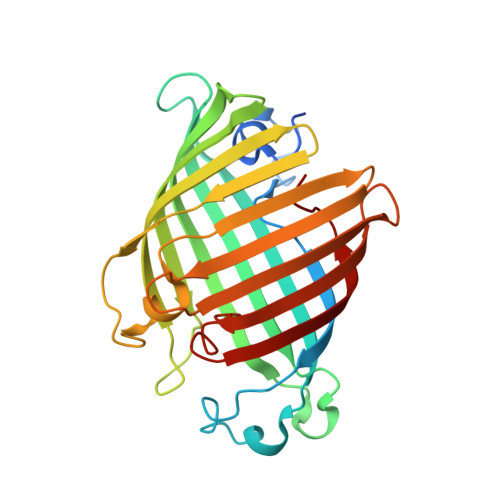
Structural evidence for dimerization-regulated activation of an integral membrane phospholipase.

Snijder, H.J.Ubarretxena-Belandia, I.Blaauw, M.Kalk, K.H.Verheij, H.M.Egmond, M.R.Dekker, N.Dijkstra, B.W.

(1999) Nature 401: 717-721

  • DOI: https://doi.org/10.1038/44890
  • Primary Citation Related Structures: 
    1QD5, 1QD6

  • PubMed Abstract: 

    Dimerization is a biological regulatory mechanism employed by both soluble and membrane proteins. However, there are few structural data on the factors that govern dimerization of membrane proteins. Outer membrane phospholipase A (OMPLA) is an integral membrane enzyme which participates in secretion of colicins in Escherichia coli. In Campilobacter and Helicobacter pylori strains, OMPLA is implied in virulence. Its activity is regulated by reversible dimerization. Here we report X-ray structures of monomeric and dimeric OMPLA from E. coli. Dimer interactions occur almost exclusively in the apolar membrane-embedded parts, with two hydrogen bonds within the hydrophobic membrane area being key interactions. Dimerization results in functional oxyanion holes and substrate-binding pockets, which are absent in monomeric OMPLA. These results provide a detailed view of activation by dimerization of a membrane protein.


  • Organizational Affiliation
    • Laboratory of Biophysical Chemistry, BIOSON Research Institute, University of Groningen, The Netherlands.

Macromolecule Content 

  • Total Structure Weight: 33 kDa 
  • Atom Count: 2,204 
  • Modeled Residue Count: 257 
  • Deposited Residue Count: 275 
  • Unique protein chains: 1

Macromolecules

Find similar proteins by:|  3D Structure
Entity ID: 1
MoleculeChains  Sequence LengthOrganismDetailsImage
OUTER MEMBRANE PHOSPHOLIPASE A275Escherichia coliMutation(s): 0 
EC: 3.1.1.32 (PDB Primary Data), 3.1.1.4 (UniProt)
Membrane Entity: Yes 
UniProt
Find proteins for P0A921 (Escherichia coli (strain K12))
Explore P0A921 
Go to UniProtKB:  P0A921
Entity Groups
Sequence Clusters30% Identity50% Identity70% Identity90% Identity95% Identity100% Identity
UniProt GroupP0A921
Sequence Annotations
Expand
Reference Sequence

Experimental Data & Validation

Experimental Data

  • Method: X-RAY DIFFRACTION
  • Resolution: 2.17 Å
  • R-Value Free:  0.270 (Depositor), 0.278 (DCC) 
  • R-Value Work:  0.221 (Depositor), 0.233 (DCC) 
  • R-Value Observed: 0.221 (Depositor) 
Space Group: P 31 2 1
Unit Cell:
Length ( Å )Angle ( ˚ )
a = 78.55α = 90
b = 78.55β = 90
c = 101.52γ = 120
Software Package:
Software NamePurpose
MLPHAREphasing
DMmodel building
X-PLORrefinement
DENZOdata reduction
XDSdata reduction
SCALEPACKdata scaling
XDSdata scaling
DMphasing

Structure Validation

View Full Validation Report


Ligand Structure Quality Assessment 


Entry History 

Deposition Data

Revision History  (Full details and data files)

  • Version 1.0: 1999-10-25
    Type: Initial release
  • Version 1.1: 2008-04-27
    Changes: Version format compliance
  • Version 1.2: 2011-07-13
    Changes: Version format compliance
  • Version 1.3: 2017-10-04
    Changes: Refinement description
  • Version 1.4: 2018-01-31
    Changes: Experimental preparation
  • Version 1.5: 2018-04-04
    Changes: Data collection
  • Version 1.6: 2018-04-11
    Changes: Data collection
  • Version 1.7: 2020-07-29
    Type: Remediation
    Reason: Carbohydrate remediation
    Changes: Data collection, Derived calculations, Structure summary
  • Version 1.8: 2024-02-14
    Changes: Data collection, Database references, Structure summary