1P77 | pdb_00001p77

CRYSTAL STRUCTURE OF SHIKIMATE DEHYDROGENASE (AROE) FROM HAEMOPHILUS INFLUENZAE


Experimental Data Snapshot

  • Method: X-RAY DIFFRACTION
  • Resolution: 1.95 Å
  • R-Value Free: 
    0.244 (Depositor) 
  • R-Value Work: 
    0.203 (Depositor) 
  • R-Value Observed: 
    0.205 (Depositor) 

Starting Model: experimental
View more details

wwPDB Validation 3D Report Full Report

Validation slider image for 1P77

This is version 2.0 of the entry. See complete history

Literature

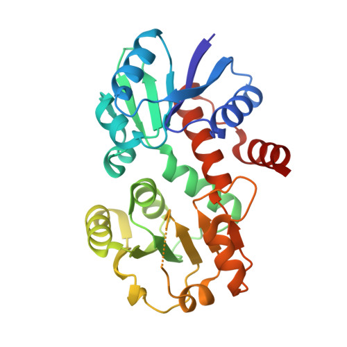
The crystal structure of shikimate dehydrogenase (AroE) reveals a unique NADPH binding mode

Ye, S.von Delft, F.Brooun, A.Knuth, M.W.Swanson, R.V.McRee, D.E.

(2003) J Bacteriol 185: 4144-4151

  • DOI: https://doi.org/10.1128/JB.185.14.4144-4151.2003
  • Primary Citation Related Structures: 
    1P74, 1P77

  • PubMed Abstract: 

    Shikimate dehydrogenase catalyzes the NADPH-dependent reversible reduction of 3-dehydroshikimate to shikimate. We report the first X-ray structure of shikimate dehydrogenase from Haemophilus influenzae to 2.4-A resolution and its complex with NADPH to 1.95-A resolution. The molecule contains two domains, a catalytic domain with a novel open twisted alpha/beta motif and an NADPH binding domain with a typical Rossmann fold. The enzyme contains a unique glycine-rich P-loop with a conserved sequence motif, GAGGXX, that results in NADPH adopting a nonstandard binding mode with the nicotinamide and ribose moieties disordered in the binary complex. A deep pocket with a narrow entrance between the two domains, containing strictly conserved residues primarily contributed by the catalytic domain, is identified as a potential 3-dehydroshikimate binding pocket. The flexibility of the nicotinamide mononucleotide portion of NADPH may be necessary for the substrate 3-dehydroshikimate to enter the pocket and for the release of the product shikimate.


  • Organizational Affiliation
    • Syrrx Inc., San Diego, California 92121, USA.

Macromolecule Content 

  • Total Structure Weight: 30.35 kDa 
  • Atom Count: 2,347 
  • Modeled Residue Count: 265 
  • Deposited Residue Count: 272 
  • Unique protein chains: 1

Macromolecules

Find similar proteins by:|  3D Structure
Entity ID: 1
MoleculeChains  Sequence LengthOrganismDetailsImage
Shikimate 5-dehydrogenase272Haemophilus influenzaeMutation(s): 0 
Gene Names: AROE OR HI0655
EC: 1.1.1.25
UniProt
Find proteins for P43876 (Haemophilus influenzae (strain ATCC 51907 / DSM 11121 / KW20 / Rd))
Explore P43876 
Go to UniProtKB:  P43876
Entity Groups
Sequence Clusters30% Identity50% Identity70% Identity90% Identity95% Identity100% Identity
UniProt GroupP43876
Sequence Annotations
Expand
Reference Sequence

Small Molecules

Ligands 2 Unique
IDChains Name / Formula / InChI Key2D Diagram3D Interactions
ATR

Query on ATR


Download:Ideal Coordinates CCD File
C [auth A]2'-MONOPHOSPHOADENOSINE-5'-DIPHOSPHATE
C10 H16 N5 O13 P3
YPTPYQSAVGGMFN-KQYNXXCUSA-N
ACT

Query on ACT


Download:Ideal Coordinates CCD File
B [auth A]ACETATE ION
C2 H3 O2
QTBSBXVTEAMEQO-UHFFFAOYSA-M

Experimental Data & Validation

Experimental Data

  • Method: X-RAY DIFFRACTION
  • Resolution: 1.95 Å
  • R-Value Free:  0.244 (Depositor) 
  • R-Value Work:  0.203 (Depositor) 
  • R-Value Observed: 0.205 (Depositor) 
Space Group: C 2 2 21
Unit Cell:
Length ( Å )Angle ( ˚ )
a = 84.054α = 90
b = 82.728β = 90
c = 82.365γ = 90
Software Package:
Software NamePurpose
REFMACrefinement
HKL-2000data reduction
SCALEPACKdata scaling
AMoREphasing

Structure Validation

View Full Validation Report


Entry History 

Deposition Data

Revision History  (Full details and data files)

  • Version 1.0: 2003-08-12
    Type: Initial release
  • Version 1.1: 2008-04-29
    Changes: Version format compliance
  • Version 1.2: 2011-07-13
    Changes: Version format compliance
  • Version 1.3: 2018-01-31
    Changes: Database references
  • Version 2.0: 2023-08-16
    Changes: Atomic model, Data collection, Database references, Derived calculations, Refinement description