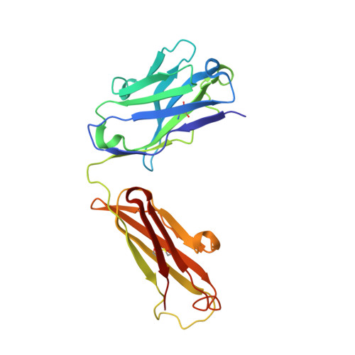
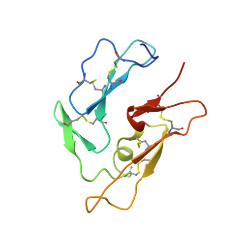
1OB1

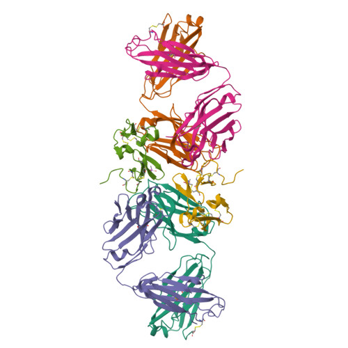
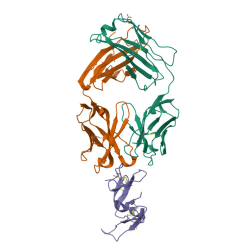
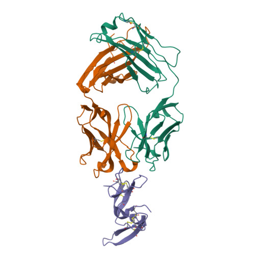
Crystal structure of a Fab complex whith Plasmodium falciparum MSP1-19


Experimental Data Snapshot

  • Method: X-RAY DIFFRACTION
  • Resolution: 2.90 Å
  • R-Value Free: 0.288 
  • R-Value Work: 0.257 
  • R-Value Observed: 0.258 

Starting Models: experimental
View more details

wwPDB Validation   3D Report Full Report


This is version 1.4 of the entry. See complete history


Literature

Crystal Structure of a Fab Complex Formed with Pfmsp1-19, the C-Terminal Fragment of Merozoite Surface Protein 1 from Plasmodium Falciparum: A Malaria Vaccine Candidate

Pizarro, J.C.Chitarra, V.Verger, D.Holm, I.Petres, S.Dartville, S.Nato, F.Longacre, S.Bentley, G.A.

(2003) J Mol Biol 328: 1091

  • DOI: https://doi.org/10.1016/s0022-2836(03)00376-0
  • Primary Citation of Related Structures:  
    1OB1

  • PubMed Abstract: 

    Merozoite surface protein 1 (MSP1) is the major protein component on the surface of the merozoite, the erythrocyte-invasive form of the malaria parasite Plasmodium. Present in all species of Plasmodium, it undergoes two distinct proteolytic maturation steps during the course of merozoite development that are essential for invasion of the erythrocyte. Antibodies specific for the C-terminal maturation product, MSP1-19, can inhibit erythrocyte invasion and parasite growth. This polypeptide is therefore considered to be one of the more promising malaria vaccine candidates. We describe here the crystal structure of recombinant MSP1-19 from P.falciparum (PfMSP1-19), the most virulent species of the parasite in humans, as a complex with the Fab fragment of the monoclonal antibody G17.12. This antibody recognises a discontinuous epitope comprising 13 residues on the first epidermal growth factor (EGF)-like domain of PfMSP1-19. Although G17.12 was raised against the recombinant antigen expressed in an insect cell/baculovirus system, it binds uniformly to the surface of merozoites from the late schizont stage, showing that the cognate epitope is exposed on the naturally occurring MSP1 polypeptide complex. Although the epitope includes residues that have been mapped to regions recognised by invasion-inhibiting antibodies studied by other workers, G17.12 does not inhibit erythrocyte invasion or MSP1 processing.


  • Organizational Affiliation

    Unité d'Immunologie Structurale (CNRS URA 2185), Département de Biologie Structurale et Chimie, Institut Pasteur, 25 rue du Dr. Roux, 75724 Paris, cedex 15, France.


Macromolecules
Find similar proteins by:  (by identity cutoff)  |  3D Structure
Entity ID: 1
MoleculeChains Sequence LengthOrganismDetailsImage
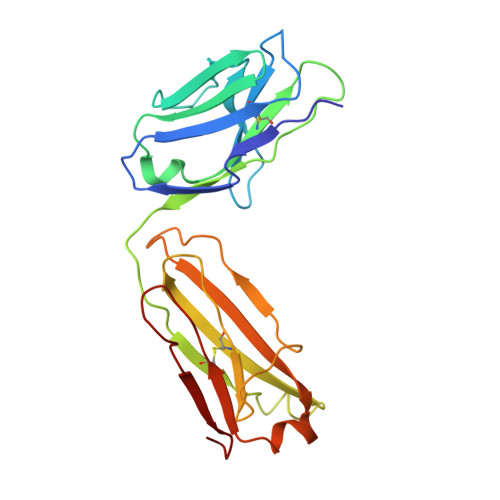
ANTIBODY, HEAVY CHAIN
A, D
215Mus musculusMutation(s): 0 
UniProt
Find proteins for A0A5E3 (Mus musculus)
Explore A0A5E3 
Go to UniProtKB:  A0A5E3
Entity Groups  
Sequence Clusters30% Identity50% Identity70% Identity90% Identity95% Identity100% Identity
UniProt GroupA0A5E3
Sequence Annotations
Expand
  • Reference Sequence
Find similar proteins by:  (by identity cutoff)  |  3D Structure
Entity ID: 2
MoleculeChains Sequence LengthOrganismDetailsImage
ANTIBODY, LIGHT CHAIN
B, E
219Mus musculusMutation(s): 0 
UniProt
Find proteins for P01863 (Mus musculus)
Explore P01863 
Go to UniProtKB:  P01863
Entity Groups  
Sequence Clusters30% Identity50% Identity70% Identity90% Identity95% Identity100% Identity
UniProt GroupP01863
Sequence Annotations
Expand
  • Reference Sequence
Find similar proteins by:  (by identity cutoff)  |  3D Structure
Entity ID: 3
MoleculeChains Sequence LengthOrganismDetailsImage
MAJOR MEROZOITE SURFACE PROTEIN
C, F
99Plasmodium falciparumMutation(s): 0 
UniProt
Find proteins for Q25976 (Plasmodium falciparum)
Explore Q25976 
Go to UniProtKB:  Q25976
Entity Groups  
Sequence Clusters30% Identity50% Identity70% Identity90% Identity95% Identity100% Identity
UniProt GroupQ25976
Sequence Annotations
Expand
  • Reference Sequence
Small Molecules
Experimental Data & Validation

Experimental Data

  • Method: X-RAY DIFFRACTION
  • Resolution: 2.90 Å
  • R-Value Free: 0.288 
  • R-Value Work: 0.257 
  • R-Value Observed: 0.258 
  • Space Group: P 1 21 1
Unit Cell:
Length ( Å )Angle ( ˚ )
a = 51.75α = 90
b = 213.46β = 100.95
c = 59.89γ = 90
Software Package:
Software NamePurpose
REFMACrefinement
DENZOdata reduction
SCALEPACKdata scaling
AMoREphasing

Structure Validation

View Full Validation Report


Entry History 

Deposition Data

Revision History  (Full details and data files)

  • Version 1.0: 2003-05-01
    Type: Initial release
  • Version 1.1: 2011-07-13
    Changes: Advisory, Version format compliance
  • Version 1.2: 2019-04-03
    Changes: Advisory, Data collection, Other, Source and taxonomy
  • Version 1.3: 2023-12-13
    Changes: Advisory, Data collection, Database references, Other, Refinement description
  • Version 1.4: 2024-10-23
    Changes: Structure summary