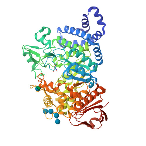
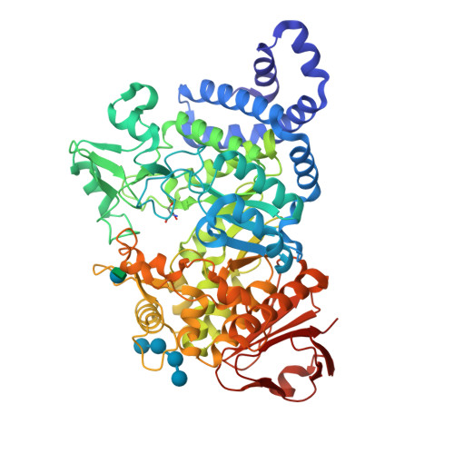
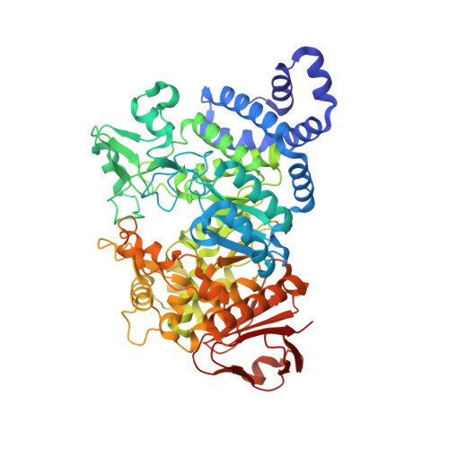
1MW2

Amylosucrase soaked with 100mM sucrose


Experimental Data Snapshot

  • Method: X-RAY DIFFRACTION
  • Resolution: 2.10 Å
  • R-Value Free: 0.226 
  • R-Value Work: 0.205 

wwPDB Validation   3D Report Full Report


This is version 2.1 of the entry. See complete history


Literature

Oligosaccharide and Sucrose Complexes of Amylosucrase. STRUCTURAL IMPLICATIONS FOR THE POLYMERASE ACTIVITY

Skov, L.K.Mirza, O.Sprogoe, D.Dar, I.Remaud-Simeon, M.Albenne, C.Monsan, P.Gajhede, M.

(2002) J Biol Chem 277: 47741-47747

  • DOI: https://doi.org/10.1074/jbc.M207860200
  • Primary Citation of Related Structures:  
    1MVY, 1MW0, 1MW1, 1MW2, 1MW3

  • PubMed Abstract: 

    The glucosyltransferase amylosucrase is structurally quite similar to the hydrolase alpha-amylase. How this switch in functionality is achieved is an important and fundamental question. The inactive E328Q amylosucrase variant has been co-crystallized with maltoheptaose, and the structure was determined by x-ray crystallography to 2.2 A resolution, revealing a maltoheptaose binding site in the B'-domain somewhat distant from the active site. Additional soaking of these crystals with maltoheptaose resulted in replacement of Tris in the active site with maltoheptaose, allowing the mapping of the -1 to +5 binding subsites. Crystals of amylosucrase were soaked with sucrose at different concentrations. The structures at approximately 2.1 A resolution revealed three new binding sites of different affinity. The highest affinity binding site is close to the active site but is not in the previously identified substrate access channel. Allosteric regulation seems necessary to facilitate access from this binding site. The structures show the pivotal role of the B'-domain in the transferase reaction. Based on these observations, an extension of the hydrolase reaction mechanism valid for this enzyme can be proposed. In this mechanism, the glycogen-like polymer is bound in the widest access channel to the active site. The polymer binding introduces structural changes that allow sucrose to migrate from its binding site into the active site and displace the polymer.


  • Organizational Affiliation

    Protein Structure Group, Department of Chemistry, University of Copenhagen, Universitetsparken 5, DK-2100 Copenhagen, Denmark. lsk@dfh.dk


Macromolecules
Find similar proteins by:  (by identity cutoff)  |  3D Structure
Entity ID: 1
MoleculeChains Sequence LengthOrganismDetailsImage
amylosucrase628Neisseria polysacchareaMutation(s): 0 
EC: 2.4.1.4
UniProt
Find proteins for Q9ZEU2 (Neisseria polysaccharea)
Explore Q9ZEU2 
Go to UniProtKB:  Q9ZEU2
Entity Groups  
Sequence Clusters30% Identity50% Identity70% Identity90% Identity95% Identity100% Identity
UniProt GroupQ9ZEU2
Sequence Annotations
Expand
  • Reference Sequence
Oligosaccharides

Help

Entity ID: 2
MoleculeChains Length2D Diagram Glycosylation3D Interactions
alpha-D-glucopyranose-(1-4)-alpha-D-glucopyranose-(1-4)-alpha-D-glucopyranose-(1-4)-alpha-D-glucopyranose
B
4N/A
Glycosylation Resources
GlyTouCan:  G87171PZ
GlyCosmos:  G87171PZ
GlyGen:  G87171PZ
Entity ID: 3
MoleculeChains Length2D Diagram Glycosylation3D Interactions
beta-D-fructofuranose-(2-1)-alpha-D-glucopyranose
C
2N/A
Glycosylation Resources
GlyTouCan:  G05551OP
GlyCosmos:  G05551OP
Small Molecules
Ligands 1 Unique
IDChains Name / Formula / InChI Key2D Diagram3D Interactions
TRS
Query on TRS

Download Ideal Coordinates CCD File 
D [auth A]2-AMINO-2-HYDROXYMETHYL-PROPANE-1,3-DIOL
C4 H12 N O3
LENZDBCJOHFCAS-UHFFFAOYSA-O
Biologically Interesting Molecules (External Reference) 2 Unique
Entity ID: 2
IDChains NameType/Class2D Diagram3D Interactions
PRD_900010
Query on PRD_900010
B
alpha-maltotetraoseOligosaccharide / Substrate analog
Entity ID: 3
IDChains NameType/Class2D Diagram3D Interactions
PRD_900003
Query on PRD_900003
C
sucroseOligosaccharide / Nutrient
Experimental Data & Validation

Experimental Data

  • Method: X-RAY DIFFRACTION
  • Resolution: 2.10 Å
  • R-Value Free: 0.226 
  • R-Value Work: 0.205 
  • Space Group: P 21 21 2
Unit Cell:
Length ( Å )Angle ( ˚ )
a = 95.617α = 90
b = 116.025β = 90
c = 60.782γ = 90
Software Package:
Software NamePurpose
MAR345data collection
SCALEPACKdata scaling
CNSrefinement
CNSphasing

Structure Validation

View Full Validation Report


Entry History 

Deposition Data

Revision History  (Full details and data files)

  • Version 1.0: 2002-12-18
    Type: Initial release
  • Version 1.1: 2008-04-28
    Changes: Version format compliance
  • Version 1.2: 2011-07-13
    Changes: Version format compliance
  • Version 1.3: 2017-10-11
    Changes: Data collection, Refinement description
  • Version 2.0: 2020-07-29
    Type: Remediation
    Reason: Carbohydrate remediation
    Changes: Atomic model, Data collection, Database references, Derived calculations, Non-polymer description, Structure summary
  • Version 2.1: 2024-03-13
    Changes: Data collection, Database references, Structure summary