1LTT

LACTOSE BINDING TO HEAT-LABILE ENTEROTOXIN REVEALED BY X-RAY CRYSTALLOGRAPHY


Experimental Data Snapshot

  • Method: X-RAY DIFFRACTION
  • Resolution: 2.30 Å
  • R-Value Work: 0.182 
  • R-Value Observed: 0.182 

wwPDB Validation   3D Report Full Report


This is version 2.1 of the entry. See complete history


Literature

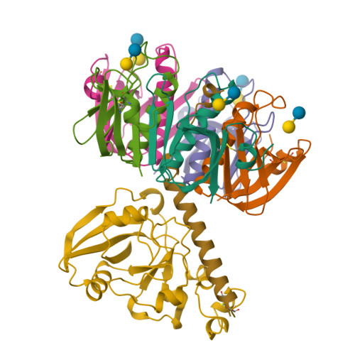
Lactose binding to heat-labile enterotoxin revealed by X-ray crystallography.

Sixma, T.K.Pronk, S.E.Kalk, K.H.van Zanten, B.A.Berghuis, A.M.Hol, W.G.

(1992) Nature 355: 561-564

  • DOI: https://doi.org/10.1038/355561a0
  • Primary Citation of Related Structures:  
    1LTT

  • PubMed Abstract: 

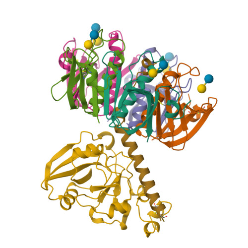
    Recognition of the oligosaccharide portion of ganglioside GM1 in membranes of target cells by the heat-labile enterotoxin from Escherichia coli is the crucial first step in its pathogenesis, as it is for the closely related cholera toxin. These toxins have five B subunits, which are essential for GM1 binding, and a single A subunit, which needs to be nicked by proteolysis and reduced, yielding an A1-'enzyme' and an A2-'linker' peptide. A1 is translocated across the membrane of intestinal epithelial cells, possibly after endocytosis, upon which it ADP-ribosylates the G protein Gs alpha. The mechanism of binding and translocation of these toxins has been extensively investigated, but how the protein is orientated on binding is still not clear. Knowing the precise arrangement of the ganglioside binding sites of the toxins will be useful for designing drugs against the diarrhoeal diseases caused by organisms secreting these toxins and in the development of oral vaccines against them. We present here the three-dimensional structure of the E. coli heat-labile enterotoxin complexed with lactose. This reveals the location of the binding site of the terminal galactose of GM1, which is consistent with toxin binding to the target cell with its A1 fragment pointing away from the membrane. A small helix is identified at the carboxy terminus of A2 which emerges through the central pore of the B subunits and probably comes into contact with the membrane upon binding, whereas the A1 subunit is flexible with respect to the B pentamer.


  • Organizational Affiliation

    BIOSON Research Institute, Department of Chemistry, University of Groningen, The Netherlands.


Macromolecules
Find similar proteins by:  (by identity cutoff)  |  3D Structure
Entity ID: 1
MoleculeChains Sequence LengthOrganismDetailsImage
HEAT-LABILE ENTEROTOXIN, SUBUNIT BA [auth D],
B [auth E],
C [auth F],
D [auth G],
E [auth H]
103Escherichia coliMutation(s): 0 
UniProt
Find proteins for P32890 (Escherichia coli)
Explore P32890 
Go to UniProtKB:  P32890
Entity Groups  
Sequence Clusters30% Identity50% Identity70% Identity90% Identity95% Identity100% Identity
UniProt GroupP32890
Sequence Annotations
Expand
  • Reference Sequence
Find similar proteins by:  (by identity cutoff)  |  3D Structure
Entity ID: 2
MoleculeChains Sequence LengthOrganismDetailsImage
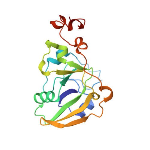
HEAT-LABILE ENTEROTOXIN, SUBUNIT AF [auth A]185Escherichia coliMutation(s): 0 
UniProt
Find proteins for P06717 (Escherichia coli)
Explore P06717 
Go to UniProtKB:  P06717
Entity Groups  
Sequence Clusters30% Identity50% Identity70% Identity90% Identity95% Identity100% Identity
UniProt GroupP06717
Sequence Annotations
Expand
  • Reference Sequence
Find similar proteins by:  (by identity cutoff)  |  3D Structure
Entity ID: 3
MoleculeChains Sequence LengthOrganismDetailsImage
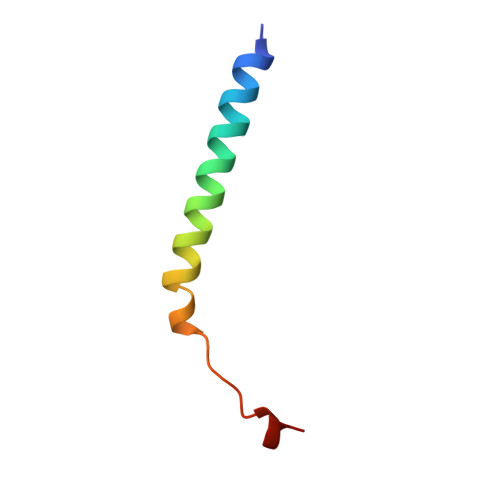
HEAT-LABILE ENTEROTOXIN, SUBUNIT AG [auth C]41Escherichia coliMutation(s): 0 
UniProt
Find proteins for P06717 (Escherichia coli)
Explore P06717 
Go to UniProtKB:  P06717
Entity Groups  
Sequence Clusters30% Identity50% Identity70% Identity90% Identity95% Identity100% Identity
UniProt GroupP06717
Sequence Annotations
Expand
  • Reference Sequence
Oligosaccharides

Help

Entity ID: 4
MoleculeChains Length2D Diagram Glycosylation3D Interactions
beta-D-galactopyranose-(1-4)-beta-D-glucopyranoseH [auth B],
I,
J,
K,
L
2N/A
Glycosylation Resources
GlyTouCan:  G84224TW
GlyCosmos:  G84224TW
GlyGen:  G84224TW
Biologically Interesting Molecules (External Reference) 1 Unique
Experimental Data & Validation

Experimental Data

  • Method: X-RAY DIFFRACTION
  • Resolution: 2.30 Å
  • R-Value Work: 0.182 
  • R-Value Observed: 0.182 
  • Space Group: P 21 21 21
Unit Cell:
Length ( Å )Angle ( ˚ )
a = 119.8α = 90
b = 101.2β = 90
c = 64.2γ = 90
Software Package:
Software NamePurpose
X-PLORmodel building
X-PLORrefinement
X-PLORphasing

Structure Validation

View Full Validation Report


Entry History 

Deposition Data

Revision History  (Full details and data files)

  • Version 1.0: 1994-01-31
    Type: Initial release
  • Version 1.1: 2008-03-24
    Changes: Version format compliance
  • Version 1.2: 2011-07-13
    Changes: Version format compliance
  • Version 2.0: 2020-07-29
    Type: Remediation
    Reason: Carbohydrate remediation
    Changes: Atomic model, Data collection, Derived calculations, Other, Structure summary
  • Version 2.1: 2024-11-20
    Changes: Data collection, Database references, Structure summary