1J7K | pdb_00001j7k

THERMOTOGA MARITIMA RUVB P216G MUTANT


Experimental Data Snapshot

  • Method: X-RAY DIFFRACTION
  • Resolution: 1.80 Å
  • R-Value Free: 
    0.238 (Depositor), 0.239 (DCC) 
  • R-Value Work: 
    0.212 (Depositor), 0.214 (DCC) 
  • R-Value Observed: 
    0.215 (Depositor) 

Starting Model: experimental
View more details

wwPDB Validation 3D Report Full Report

Validation slider image for 1J7K

Ligand Structure Quality Assessment 


This is version 1.4 of the entry. See complete history

Literature

Structure and mechanism of the RuvB Holliday junction branch migration motor.

Putnam, C.D.Clancy, S.B.Tsuruta, H.Gonzalez, S.Wetmur, J.G.Tainer, J.A.

(2001) J Mol Biology 311: 297-310

  • DOI: https://doi.org/10.1006/jmbi.2001.4852
  • Primary Citation Related Structures: 
    1IN4, 1IN5, 1IN6, 1IN7, 1IN8, 1J7K

  • PubMed Abstract: 

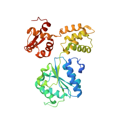
    The RuvB hexamer is the chemomechanical motor of the RuvAB complex that migrates Holliday junction branch-points in DNA recombination and the rescue of stalled DNA replication forks. The 1.6 A crystal structure of Thermotoga maritima RuvB together with five mutant structures reveal that RuvB is an ATPase-associated with diverse cellular activities (AAA+-class ATPase) with a winged-helix DNA-binding domain. The RuvB-ADP complex structure and mutagenesis suggest how AAA+-class ATPases couple nucleotide binding and hydrolysis to interdomain conformational changes and asymmetry within the RuvB hexamer implied by the crystallographic packing and small-angle X-ray scattering in solution. ATP-driven domain motion is positioned to move double-stranded DNA through the hexamer and drive conformational changes between subunits by altering the complementary hydrophilic protein- protein interfaces. Structural and biochemical analysis of five motifs in the protein suggest that ATP binding is a strained conformation recognized both by sensors and the Walker motifs and that intersubunit activation occurs by an arginine finger motif reminiscent of the GTPase-activating proteins. Taken together, these results provide insights into how RuvB functions as a motor for branch migration of Holliday junctions.


  • Organizational Affiliation
    • Department of Molecular Biology, Skaggs Institute for Chemical Biology, The Scripps Research Institute, MB 4, 10550 North Torrey Pines Rd, La Jolla, CA 92037, USA.

Macromolecule Content 

  • Total Structure Weight: 37.97 kDa 
  • Atom Count: 2,680 
  • Modeled Residue Count: 299 
  • Deposited Residue Count: 334 
  • Unique protein chains: 1

Macromolecules

Find similar proteins by:|  3D Structure
Entity ID: 1
MoleculeChains  Sequence LengthOrganismDetailsImage
HOLLIDAY JUNCTION DNA HELICASE RUVB334Thermotoga maritimaMutation(s): 1 
Gene Names: RuvB
EC: 3.6.4
UniProt
Find proteins for Q56313 (Thermotoga maritima (strain ATCC 43589 / DSM 3109 / JCM 10099 / NBRC 100826 / MSB8))
Explore Q56313 
Go to UniProtKB:  Q56313
Entity Groups
Sequence Clusters30% Identity50% Identity70% Identity90% Identity95% Identity100% Identity
UniProt GroupQ56313
Sequence Annotations
Expand
Reference Sequence

Experimental Data & Validation

Experimental Data

  • Method: X-RAY DIFFRACTION
  • Resolution: 1.80 Å
  • R-Value Free:  0.238 (Depositor), 0.239 (DCC) 
  • R-Value Work:  0.212 (Depositor), 0.214 (DCC) 
  • R-Value Observed: 0.215 (Depositor) 
Space Group: P 65
Unit Cell:
Length ( Å )Angle ( ˚ )
a = 86.217α = 90
b = 86.217β = 90
c = 82.142γ = 120
Software Package:
Software NamePurpose
AMoREphasing
CNSrefinement
DENZOdata reduction
SCALEPACKdata scaling

Structure Validation

View Full Validation Report


Ligand Structure Quality Assessment 


Entry History 

Deposition Data

Revision History  (Full details and data files)

  • Version 1.0: 2001-08-08
    Type: Initial release
  • Version 1.1: 2008-04-27
    Changes: Version format compliance
  • Version 1.2: 2011-07-13
    Changes: Version format compliance
  • Version 1.3: 2021-10-27
    Changes: Database references, Derived calculations
  • Version 1.4: 2023-08-16
    Changes: Data collection, Refinement description