1G0D | pdb_00001g0d

CRYSTAL STRUCTURE OF RED SEA BREAM TRANSGLUTAMINASE


Experimental Data Snapshot

  • Method: X-RAY DIFFRACTION
  • Resolution: 2.50 Å
  • R-Value Free: 
    0.249 (Depositor) 
  • R-Value Work: 
    0.200 (Depositor), 0.196 (DCC) 

wwPDB Validation 3D Report Full Report

Validation slider image for 1G0D

This is version 1.3 of the entry. See complete history

Literature

Crystal structure of red sea bream transglutaminase.

Noguchi, K.Ishikawa, K.Yokoyama, K.i.Ohtsuka, T.Nio, N.Suzuki, E.

(2001) J Biological Chem 276: 12055-12059

  • DOI: https://doi.org/10.1074/jbc.M009862200
  • Primary Citation Related Structures: 
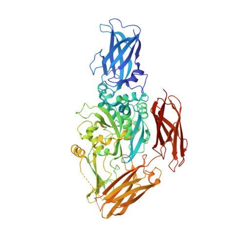
    1G0D

  • PubMed Abstract: 

    The crystal structure of the tissue-type transglutaminase from red sea bream liver (fish-derived transglutaminase, FTG) has been determined at 2.5-A resolution using the molecular replacement method, based on the crystal structure of human blood coagulation factor XIII, which is a transglutaminase zymogen. The model contains 666 residues of a total of 695 residues, 382 water molecules, and 1 sulfate ion. FTG consists of four domains, and its overall and active site structures are similar to those of human factor XIII. However, significant structural differences are observed in both the acyl donor and acyl acceptor binding sites, which account for the difference in substrate preferences. The active site of the enzyme is inaccessible to the solvent, because the catalytic Cys-272 hydrogen-bonds to Tyr-515, which is thought to be displaced upon acyl donor binding to FTG. It is postulated that the binding of an inappropriate substrate to FTG would lead to inactivation of the enzyme because of the formation of a new disulfide bridge between Cys-272 and the adjacent Cys-333 immediately after the displacement of Tyr-515. Considering the mutational studies previously reported on the tissue-type transglutaminases, we propose that Cys-333 and Tyr-515 are important in strictly controlling the enzymatic activity of FTG.


  • Organizational Affiliation
    • Central Research Laboratories and Food Research & Development Laboratories, Ajinomoto Company Inc., 1-1 Suzuki-cho, Kawasaki-ku, Kawasaki, Kanagawa 210-8681, Japan.

Macromolecule Content 

  • Total Structure Weight: 78.42 kDa 
  • Atom Count: 5,671 
  • Modeled Residue Count: 666 
  • Deposited Residue Count: 695 
  • Unique protein chains: 1

Macromolecules

Find similar proteins by:|  3D Structure
Entity ID: 1
MoleculeChains  Sequence LengthOrganismDetailsImage
PROTEIN-GLUTAMINE GAMMA-GLUTAMYLTRANSFERASE695Pagrus majorMutation(s): 0 
EC: 2.3.2.13 (PDB Primary Data), 2.3.1 (UniProt), 3.4 (UniProt), 3.5.1.44 (UniProt)
UniProt
Find proteins for P52181 (Pagrus major)
Explore P52181 
Go to UniProtKB:  P52181
Entity Groups
Sequence Clusters30% Identity50% Identity70% Identity90% Identity95% Identity100% Identity
UniProt GroupP52181
Sequence Annotations
Expand
Reference Sequence

Small Molecules

Ligands 1 Unique
IDChains Name / Formula / InChI Key2D Diagram3D Interactions
SO4

Query on SO4


Download:Ideal Coordinates CCD File
B [auth A]SULFATE ION
O4 S
QAOWNCQODCNURD-UHFFFAOYSA-L

Experimental Data & Validation

Experimental Data

  • Method: X-RAY DIFFRACTION
  • Resolution: 2.50 Å
  • R-Value Free:  0.249 (Depositor) 
  • R-Value Work:  0.200 (Depositor), 0.196 (DCC) 
Space Group: P 65 2 2
Unit Cell:
Length ( Å )Angle ( ˚ )
a = 97.8α = 90
b = 97.8β = 90
c = 455.5γ = 120
Software Package:
Software NamePurpose
AMoREphasing
X-PLORrefinement
DENZOdata reduction
SCALEPACKdata scaling

Structure Validation

View Full Validation Report


Entry History 

Deposition Data

Revision History  (Full details and data files)

  • Version 1.0: 2001-05-23
    Type: Initial release
  • Version 1.1: 2008-04-27
    Changes: Version format compliance
  • Version 1.2: 2011-07-13
    Changes: Version format compliance
  • Version 1.3: 2024-03-13
    Changes: Data collection, Database references, Derived calculations