1FZC | pdb_00001fzc

CRYSTAL STRUCTURE OF FRAGMENT DOUBLE-D FROM HUMAN FIBRIN WITH TWO DIFFERENT BOUND LIGANDS


Experimental Data Snapshot

  • Method: X-RAY DIFFRACTION
  • Resolution: 2.30 Å
  • R-Value Free: 
    0.290 (Depositor) 
  • R-Value Work: 
    0.220 (Depositor) 

Starting Model: experimental
View more details

wwPDB Validation 3D Report Full Report

Validation slider image for 1FZC

This is version 3.0 of the entry. See complete history

Literature

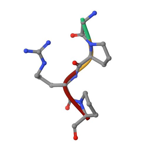
Crystal structure of fragment double-D from human fibrin with two different bound ligands.

Everse, S.J.Spraggon, G.Veerapandian, L.Riley, M.Doolittle, R.F.

(1998) Biochemistry 37: 8637-8642

  • DOI: https://doi.org/10.1021/bi9804129
  • Primary Citation Related Structures: 
    1FZC

  • PubMed Abstract: 

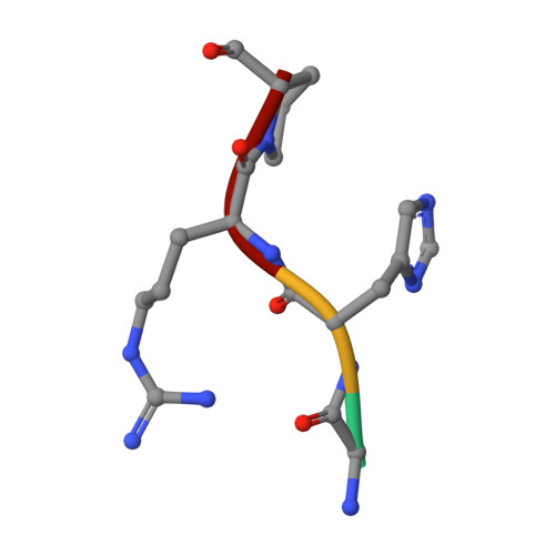
    Factor XIII-cross-linked fragment D (double-D) from human fibrin was crystallized in the presence of two different peptide ligands and the X-ray structure determined at 2.3 A. The peptide Gly-Pro-Arg-Pro-amide, which is an analogue of the knob exposed by the thrombin-catalyzed removal of fibrinopeptide A, was found to reside in the gamma-chain holes, and the peptide Gly-His-Arg-Pro-amide, which corresponds to the beta-chain knob, was found in the homologous beta-chain holes. The structure shows for the first time that the beta-chain knob does indeed bind to a homologous hole on the beta-chain. The gamma- and beta-chain holes are structurally very similar, and it is remarkable they are able to distinguish between these two peptides that differ by a single amino acid. Additionally, we have found that the beta-chain domain, like its gamma-chain counterpart, binds calcium.


  • Organizational Affiliation
    • Center for Molecular Genetics, University of California, San Diego, La Jolla, California 92093-0634, USA.

Macromolecule Content 

  • Total Structure Weight: 171.32 kDa 
  • Atom Count: 11,594 
  • Modeled Residue Count: 1,382 
  • Deposited Residue Count: 1,484 
  • Unique protein chains: 5

Macromolecules

Find similar proteins by:|  3D Structure
Entity ID: 1
MoleculeChains  Sequence LengthOrganismDetailsImage
FIBRIN
A, D
87Homo sapiensMutation(s): 0 
UniProt & NIH Common Fund Data Resources
Find proteins for P02671 (Homo sapiens)
Explore P02671 
Go to UniProtKB:  P02671
PHAROS:  P02671
GTEx:  ENSG00000171560 
Entity Groups
Sequence Clusters30% Identity50% Identity70% Identity90% Identity95% Identity100% Identity
UniProt GroupP02671
Sequence Annotations
Expand
Reference Sequence
Find similar proteins by:|  3D Structure
Entity ID: 2
MoleculeChains  Sequence LengthOrganismDetailsImage
FIBRIN
B, E
328Homo sapiensMutation(s): 0 
UniProt & NIH Common Fund Data Resources
Find proteins for P02675 (Homo sapiens)
Explore P02675 
Go to UniProtKB:  P02675
PHAROS:  P02675
GTEx:  ENSG00000171564 
Entity Groups
Sequence Clusters30% Identity50% Identity70% Identity90% Identity95% Identity100% Identity
UniProt GroupP02675
Glycosylation
Glycosylation Sites: 1Go to GlyGen: P02675-1
Sequence Annotations
Expand
Reference Sequence
Find similar proteins by:|  3D Structure
Entity ID: 3
MoleculeChains  Sequence LengthOrganismDetailsImage
FIBRIN
C, F
319Homo sapiensMutation(s): 0 
UniProt & NIH Common Fund Data Resources
Find proteins for P02679 (Homo sapiens)
Explore P02679 
Go to UniProtKB:  P02679
PHAROS:  P02679
GTEx:  ENSG00000171557 
Entity Groups
Sequence Clusters30% Identity50% Identity70% Identity90% Identity95% Identity100% Identity
UniProt GroupP02679
Sequence Annotations
Expand
Reference Sequence
Find similar proteins by:  Sequence   |   3D Structure  
Entity ID: 4
MoleculeChains  Sequence LengthOrganismDetailsImage
FIBRIN
G, H
4Homo sapiensMutation(s): 0 
Find similar proteins by:  Sequence   |   3D Structure  
Entity ID: 5
MoleculeChains  Sequence LengthOrganismDetailsImage
FIBRIN
I, J
4Homo sapiensMutation(s): 0 

Oligosaccharides

Help  
Entity ID: 6
MoleculeChains Length2D Diagram GlycosylationD Interactions
2-acetamido-2-deoxy-beta-D-glucopyranose-(1-4)-2-acetamido-2-deoxy-beta-D-glucopyranose
K
2N-Glycosylation
Glycosylation Resources
GlyTouCan: G42666HT
GlyCosmos: G42666HT
GlyGen: G42666HT

Experimental Data & Validation

Experimental Data

  • Method: X-RAY DIFFRACTION
  • Resolution: 2.30 Å
  • R-Value Free:  0.290 (Depositor) 
  • R-Value Work:  0.220 (Depositor) 
Space Group: P 1 21 1
Unit Cell:
Length ( Å )Angle ( ˚ )
a = 83.443α = 90
b = 95.6β = 90.19
c = 113.641γ = 90
Software Package:
Software NamePurpose
DENZOdata reduction
SCALEPACKdata scaling
X-PLORmodel building
X-PLORrefinement
X-PLORphasing

Structure Validation

View Full Validation Report


Entry History 

Deposition Data

Revision History  (Full details and data files)

  • Version 1.0: 1998-10-14
    Type: Initial release
  • Version 1.1: 2008-03-10
    Changes: Version format compliance
  • Version 1.2: 2011-07-13
    Changes: Non-polymer description, Version format compliance
  • Version 2.0: 2020-07-29
    Type: Remediation
    Reason: Carbohydrate remediation
    Changes: Advisory, Atomic model, Data collection, Database references, Derived calculations, Structure summary
  • Version 2.1: 2024-04-03
    Changes: Data collection, Database references, Refinement description, Structure summary
  • Version 3.0: 2024-12-25
    Changes: Advisory, Atomic model, Data collection, Derived calculations, Structure summary