1F8R | pdb_00001f8r

CRYSTAL STRUCTURE OF L-AMINO ACID OXIDASE FROM CALLOSELASMA RHODOSTOMA COMPLEXED WITH CITRATE


Experimental Data Snapshot

  • Method: X-RAY DIFFRACTION
  • Resolution: 2.00 Å
  • R-Value Free: 
    0.210 (Depositor), 0.211 (DCC) 
  • R-Value Work: 
    0.185 (Depositor), 0.186 (DCC) 

wwPDB Validation 3D Report Full Report

Validation slider image for 1F8R

Ligand Structure Quality Assessment 


This is version 2.1 of the entry. See complete history

Literature

The structure of L-amino acid oxidase reveals the substrate trajectory into an enantiomerically conserved active site.

Pawelek, P.D.Cheah, J.Coulombe, R.Macheroux, P.Ghisla, S.Vrielink, A.

(2000) EMBO J 19: 4204-4215

  • DOI: https://doi.org/10.1093/emboj/19.16.4204
  • Primary Citation Related Structures: 
    1F8R, 1F8S

  • PubMed Abstract: 

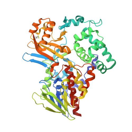
    The structure of L-amino acid oxidase (LAAO) from Calloselasma rhodostoma has been determined to 2.0 A resolution in the presence of two ligands: citrate and o-aminobenzoate (AB). The protomer consists of three domains: an FAD-binding domain, a substrate-binding domain and a helical domain. The interface between the substrate-binding and helical domains forms a 25 A long funnel, which provides access to the active site. Three AB molecules are visible within the funnel of the LAAO-AB complex; their orientations suggest the trajectory of the substrate to the active site. The innermost AB molecule makes hydrogen bond contacts with the active site residues, Arg90 and Gly464, and the aromatic portion of the ligand is situated in a hydrophobic pocket. These contacts are proposed to mimic those of the natural substrate. Comparison of LAAO with the structure of mammalian D-amino acid oxidase reveals significant differences in their modes of substrate entry. Furthermore, a mirror-symmetrical relationship between the two substrate-binding sites is observed which facilitates enantiomeric selectivity while preserving a common arrangement of the atoms involved in catalysis.


  • Organizational Affiliation
    • Biochemistry Department and Montréal Joint Center for Structural Biology, McIntyre Medical Sciences Building, McGill University, 3655 Promenade Sir William Osler, Montréal, Québec, H3G 1Y6, Canada.

Macromolecule Content 

  • Total Structure Weight: 232.27 kDa 
  • Atom Count: 17,632 
  • Modeled Residue Count: 1,932 
  • Deposited Residue Count: 1,992 
  • Unique protein chains: 1

Macromolecules

Find similar proteins by:|  3D Structure
Entity ID: 1
MoleculeChains  Sequence LengthOrganismDetailsImage
L-AMINO ACID OXIDASE
A, B, C, D
498Calloselasma rhodostomaMutation(s): 0 
EC: 1.4.3.2
UniProt
Find proteins for P81382 (Calloselasma rhodostoma)
Explore P81382 
Go to UniProtKB:  P81382
Entity Groups
Sequence Clusters30% Identity50% Identity70% Identity90% Identity95% Identity100% Identity
UniProt GroupP81382
Glycosylation
Glycosylation Sites: 2
Sequence Annotations
Expand
Reference Sequence

Oligosaccharides

Help  
Entity ID: 2
MoleculeChains Length2D Diagram GlycosylationD Interactions
2-acetamido-2-deoxy-beta-D-glucopyranose-(1-4)-[alpha-L-fucopyranose-(1-6)]2-acetamido-2-deoxy-beta-D-glucopyranose
E, F, G, H
3N-Glycosylation
Glycosylation Resources
GlyTouCan: G21290RB
GlyCosmos: G21290RB
GlyGen: G21290RB

Small Molecules

Ligands 3 Unique
IDChains Name / Formula / InChI Key2D Diagram3D Interactions
FAD

Query on FAD


Download:Ideal Coordinates CCD File
K [auth A],
N [auth B],
Q [auth C],
T [auth D]
FLAVIN-ADENINE DINUCLEOTIDE
C27 H33 N9 O15 P2
VWWQXMAJTJZDQX-UYBVJOGSSA-N
NAG

Query on NAG


Download:Ideal Coordinates CCD File
I [auth A],
L [auth B],
O [auth C],
R [auth D]
2-acetamido-2-deoxy-beta-D-glucopyranose
C8 H15 N O6
OVRNDRQMDRJTHS-FMDGEEDCSA-N
CIT

Query on CIT


Download:Ideal Coordinates CCD File
J [auth A],
M [auth B],
P [auth C],
S [auth D]
CITRIC ACID
C6 H8 O7
KRKNYBCHXYNGOX-UHFFFAOYSA-N

Experimental Data & Validation

Experimental Data

  • Method: X-RAY DIFFRACTION
  • Resolution: 2.00 Å
  • R-Value Free:  0.210 (Depositor), 0.211 (DCC) 
  • R-Value Work:  0.185 (Depositor), 0.186 (DCC) 
Space Group: P 1 21 1
Unit Cell:
Length ( Å )Angle ( ˚ )
a = 79.496α = 90
b = 154.962β = 109.49
c = 102.947γ = 90
Software Package:
Software NamePurpose
DENZOdata reduction
SCALEPACKdata scaling
SHARPphasing
CNSrefinement

Structure Validation

View Full Validation Report


Ligand Structure Quality Assessment 


Entry History 

Deposition Data

Revision History  (Full details and data files)

  • Version 1.0: 2000-08-24
    Type: Initial release
  • Version 1.1: 2008-04-27
    Changes: Version format compliance
  • Version 1.2: 2011-07-13
    Changes: Non-polymer description, Version format compliance
  • Version 2.0: 2020-07-29
    Type: Remediation
    Reason: Carbohydrate remediation
    Changes: Advisory, Atomic model, Data collection, Derived calculations, Structure summary
  • Version 2.1: 2024-11-13
    Changes: Data collection, Database references, Structure summary