1EAX | pdb_00001eax

Crystal structure of MTSP1 (matriptase)


Experimental Data Snapshot

  • Method: X-RAY DIFFRACTION
  • Resolution: 1.30 Å
  • R-Value Free: 
    0.193 (Depositor) 
  • R-Value Work: 
    0.184 (Depositor) 
  • R-Value Observed: 
    0.184 (Depositor) 

Starting Model: experimental
View more details

wwPDB Validation 3D Report Full Report

Validation slider image for 1EAX

This is version 1.3 of the entry. See complete history

Literature

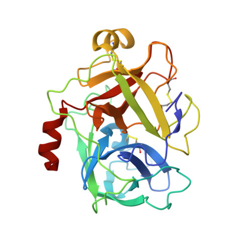
Catalytic Domain Structures of Mt-Sp1/Matriptase, a Matrix-Degrading Transmembrane Serine Proteinase.

Friedrich, R.Fuentes-Prior, P.Ong, E.Coombs, G.Hunter, M.Oehler, R.Pierson, D.Gonzalez, R.Huber, R.Bode, W.Madison, E.L.

(2002) J Biological Chem 277: 2160

  • DOI: https://doi.org/10.1074/jbc.M109830200
  • Primary Citation Related Structures: 
    1EAW, 1EAX

  • PubMed Abstract: 

    The type II transmembrane multidomain serine proteinase MT-SP1/matriptase is highly expressed in many human cancer-derived cell lines and has been implicated in extracellular matrix re-modeling, tumor growth, and metastasis. We have expressed the catalytic domain of MT-SP1 and solved the crystal structures of complexes with benzamidine at 1.3 A and bovine pancreatic trypsin inhibitor at 2.9 A. MT-SP1 exhibits a trypsin-like serine proteinase fold, featuring a unique nine-residue 60-insertion loop that influences interactions with protein substrates. The structure discloses a trypsin-like S1 pocket, a small hydrophobic S2 subsite, and an open negatively charged S4 cavity that favors the binding of basic P3/P4 residues. A complementary charge pattern on the surface opposite the active site cleft suggests a distinct docking of the preceding low density lipoprotein receptor class A domain. The benzamidine crystals possess a freely accessible active site and are hence well suited for soaking small molecules, facilitating the improvement of inhibitors. The crystal structure of the MT-SP1 complex with bovine pancreatic trypsin inhibitor serves as a model for hepatocyte growth factor activator inhibitor 1, the physiological inhibitor of MT-SP1, and suggests determinants for the substrate specificity.


  • Organizational Affiliation
    • Max-Planck-Institut für Biochemie, Abteilung Strukturforschung, Am Klopferspitz 18a, 82152 Martinsried, Germany.

Macromolecule Content 

  • Total Structure Weight: 26.68 kDa 
  • Atom Count: 2,259 
  • Modeled Residue Count: 241 
  • Deposited Residue Count: 241 
  • Unique protein chains: 1

Macromolecules

Find similar proteins by:|  3D Structure
Entity ID: 1
MoleculeChains  Sequence LengthOrganismDetailsImage
SUPPRESSOR OF TUMORIGENICITY 14241Homo sapiensMutation(s): 0 
EC: 3.4.21 (PDB Primary Data), 3.4.21.109 (UniProt)
UniProt & NIH Common Fund Data Resources
Find proteins for Q9Y5Y6 (Homo sapiens)
Explore Q9Y5Y6 
Go to UniProtKB:  Q9Y5Y6
PHAROS:  Q9Y5Y6
GTEx:  ENSG00000149418 
Entity Groups
Sequence Clusters30% Identity50% Identity70% Identity90% Identity95% Identity100% Identity
UniProt GroupQ9Y5Y6
Sequence Annotations
Expand
Reference Sequence

Small Molecules

Ligands 2 Unique
IDChains Name / Formula / InChI Key2D Diagram3D Interactions
BEN

Query on BEN


Download:Ideal Coordinates CCD File
C [auth A]BENZAMIDINE
C7 H8 N2
PXXJHWLDUBFPOL-UHFFFAOYSA-N
SO4

Query on SO4


Download:Ideal Coordinates CCD File
B [auth A]SULFATE ION
O4 S
QAOWNCQODCNURD-UHFFFAOYSA-L

Experimental Data & Validation

Experimental Data

  • Method: X-RAY DIFFRACTION
  • Resolution: 1.30 Å
  • R-Value Free:  0.193 (Depositor) 
  • R-Value Work:  0.184 (Depositor) 
  • R-Value Observed: 0.184 (Depositor) 
Space Group: C 2 2 2
Unit Cell:
Length ( Å )Angle ( ˚ )
a = 66.918α = 90
b = 141.604β = 90
c = 51.936γ = 90
Software Package:
Software NamePurpose
CNSrefinement
MOSFLMdata reduction
SCALAdata scaling
AMoREphasing

Structure Validation

View Full Validation Report


Entry History 

Deposition Data

Revision History  (Full details and data files)

  • Version 1.0: 2002-01-28
    Type: Initial release
  • Version 1.1: 2015-04-15
    Changes: Database references, Derived calculations, Non-polymer description, Other, Refinement description, Version format compliance
  • Version 1.2: 2023-12-13
    Changes: Data collection, Database references, Derived calculations, Refinement description
  • Version 1.3: 2024-10-09
    Changes: Structure summary