1EAW

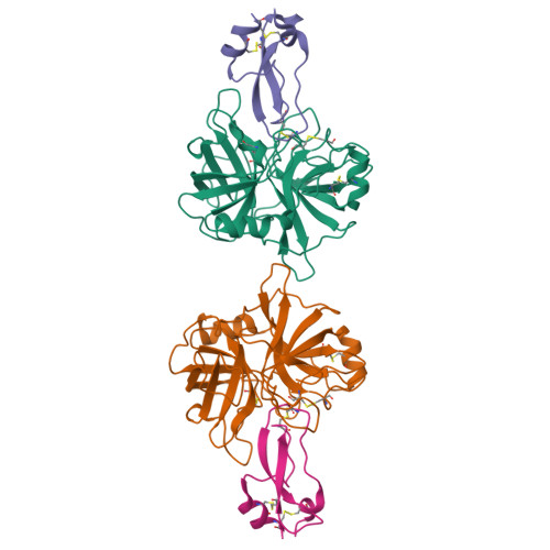
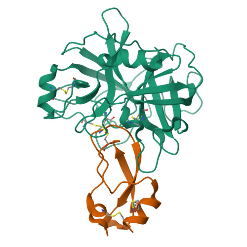
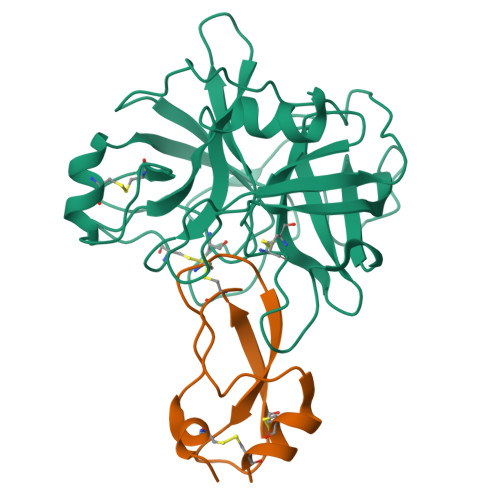
Crystal structure of the MTSP1 (matriptase)-BPTI (aprotinin) complex


Experimental Data Snapshot

  • Method: X-RAY DIFFRACTION
  • Resolution: 2.93 Å
  • R-Value Free: 0.279 
  • R-Value Work: 0.199 
  • R-Value Observed: 0.199 

Starting Model: experimental
View more details

wwPDB Validation   3D Report Full Report


This is version 1.3 of the entry. See complete history


Literature

Catalytic Domain Structures of Mt-Sp1/Matriptase, a Matrix-Degrading Transmembrane Serine Proteinase.

Friedrich, R.Fuentes-Prior, P.Ong, E.Coombs, G.Hunter, M.Oehler, R.Pierson, D.Gonzalez, R.Huber, R.Bode, W.Madison, E.L.

(2002) J Biol Chem 277: 2160

  • DOI: https://doi.org/10.1074/jbc.M109830200
  • Primary Citation of Related Structures:  
    1EAW, 1EAX

  • PubMed Abstract: 

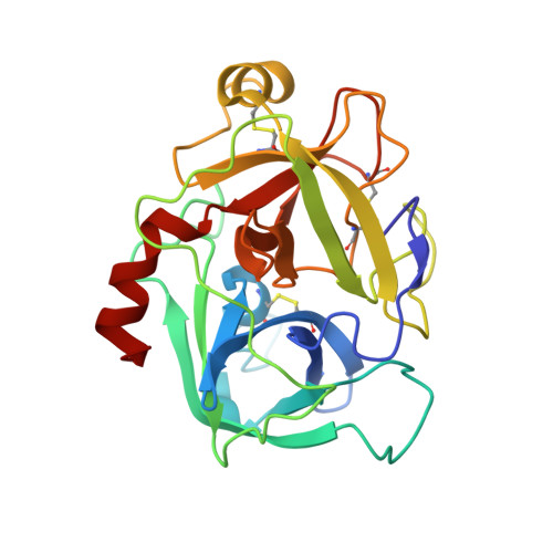
    The type II transmembrane multidomain serine proteinase MT-SP1/matriptase is highly expressed in many human cancer-derived cell lines and has been implicated in extracellular matrix re-modeling, tumor growth, and metastasis. We have expressed the catalytic domain of MT-SP1 and solved the crystal structures of complexes with benzamidine at 1.3 A and bovine pancreatic trypsin inhibitor at 2.9 A. MT-SP1 exhibits a trypsin-like serine proteinase fold, featuring a unique nine-residue 60-insertion loop that influences interactions with protein substrates. The structure discloses a trypsin-like S1 pocket, a small hydrophobic S2 subsite, and an open negatively charged S4 cavity that favors the binding of basic P3/P4 residues. A complementary charge pattern on the surface opposite the active site cleft suggests a distinct docking of the preceding low density lipoprotein receptor class A domain. The benzamidine crystals possess a freely accessible active site and are hence well suited for soaking small molecules, facilitating the improvement of inhibitors. The crystal structure of the MT-SP1 complex with bovine pancreatic trypsin inhibitor serves as a model for hepatocyte growth factor activator inhibitor 1, the physiological inhibitor of MT-SP1, and suggests determinants for the substrate specificity.


  • Organizational Affiliation

    Max-Planck-Institut für Biochemie, Abteilung Strukturforschung, Am Klopferspitz 18a, 82152 Martinsried, Germany.


Macromolecules
Find similar proteins by:  (by identity cutoff)  |  3D Structure
Entity ID: 1
MoleculeChains Sequence LengthOrganismDetailsImage
SUPPRESSOR OF TUMORIGENICITY 14
A, C
241Homo sapiensMutation(s): 0 
EC: 3.4.21 (PDB Primary Data), 3.4.21.109 (UniProt)
UniProt & NIH Common Fund Data Resources
Find proteins for Q9Y5Y6 (Homo sapiens)
Explore Q9Y5Y6 
Go to UniProtKB:  Q9Y5Y6
PHAROS:  Q9Y5Y6
GTEx:  ENSG00000149418 
Entity Groups  
Sequence Clusters30% Identity50% Identity70% Identity90% Identity95% Identity100% Identity
UniProt GroupQ9Y5Y6
Sequence Annotations
Expand
  • Reference Sequence
Find similar proteins by:  (by identity cutoff)  |  3D Structure
Entity ID: 2
MoleculeChains Sequence LengthOrganismDetailsImage
PANCREATIC TRYPSIN INHIBITOR
B, D
58Bos taurusMutation(s): 0 
UniProt
Find proteins for P00974 (Bos taurus)
Explore P00974 
Go to UniProtKB:  P00974
Entity Groups  
Sequence Clusters30% Identity50% Identity70% Identity90% Identity95% Identity100% Identity
UniProt GroupP00974
Sequence Annotations
Expand
  • Reference Sequence
Experimental Data & Validation

Experimental Data

  • Method: X-RAY DIFFRACTION
  • Resolution: 2.93 Å
  • R-Value Free: 0.279 
  • R-Value Work: 0.199 
  • R-Value Observed: 0.199 
  • Space Group: P 1
Unit Cell:
Length ( Å )Angle ( ˚ )
a = 47.099α = 107.62
b = 54.233β = 96.86
c = 67.824γ = 103.36
Software Package:
Software NamePurpose
CNSrefinement
MOSFLMdata reduction
SCALAdata scaling
AMoREphasing

Structure Validation

View Full Validation Report


Entry History 

Deposition Data

Revision History  (Full details and data files)

  • Version 1.0: 2002-01-28
    Type: Initial release
  • Version 1.1: 2015-04-01
    Changes: Data collection, Database references, Derived calculations, Non-polymer description, Other, Refinement description, Version format compliance
  • Version 1.2: 2023-12-13
    Changes: Data collection, Database references, Refinement description
  • Version 1.3: 2024-11-13
    Changes: Structure summary