1E0R | pdb_00001e0r

Beta-apical domain of thermosome


Experimental Data Snapshot

  • Method: X-RAY DIFFRACTION
  • Resolution: 2.80 Å
  • R-Value Free: 
    0.318 (Depositor), 0.305 (DCC) 
  • R-Value Work: 
    0.261 (Depositor), 0.270 (DCC) 
  • R-Value Observed: 
    0.261 (Depositor) 

Starting Model: experimental
View more details

wwPDB Validation 3D Report Full Report

Validation slider image for 1E0R

This is version 1.4 of the entry. See complete history

Literature

Crystal Structure of the Beta-Apical Domain from Thermosome Reveals Structural Plasticity in Protrusion Region

Bosch, G.Baumeister, W.Essen, L.-O.

(2000) J Mol Biology 301: 19

  • DOI: https://doi.org/10.1006/jmbi.2000.3955
  • Primary Citation Related Structures: 
    1E0R

  • PubMed Abstract: 

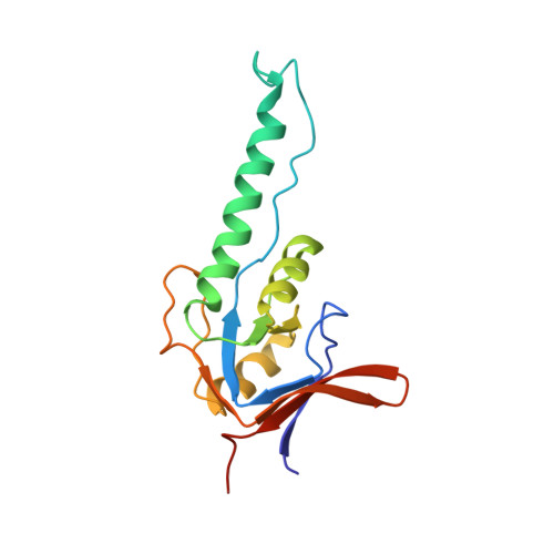
    The crystal structure of the beta-apical domain of the thermosome, an archaeal group II chaperonin from Thermoplasma acidophilum, has been determined at 2.8 A resolution. The structure shows an invariant globular core from which a 25 A long protrusion emanates, composed of an elongated alpha-helix (H10) and a long extended stretch consisting of residues GluB245-ThrB253. A comparison with previous apical domain structures reveals a large segmental displacement of the protruding part of helix H10 via the hinge GluB276-ValB278. The region comprising residues GluB245-ThrB253 adopts an extended beta-like conformation rather than the alpha-helix seen in the alpha-apical domain. Consequently, it appears that the protrusions of the apical domains from group II chaperonins might assume a variety of context-dependent conformations during an open, substrate-accepting state of the chaperonin. Sequence variations in the protrusion regions that are found in the eukaryotic TRiC/CCT subunits may provide different structural propensities and hence serve different roles in substrate recognition.


  • Organizational Affiliation
    • Max-Planck-Institute for Biochemistry, Am Klopferspitz 18a, Martinsried bei München, D-82152, Germany.

Macromolecule Content 

  • Total Structure Weight: 17.76 kDa 
  • Atom Count: 1,160 
  • Modeled Residue Count: 154 
  • Deposited Residue Count: 159 
  • Unique protein chains: 1

Macromolecules

Find similar proteins by:|  3D Structure
Entity ID: 1
MoleculeChains  Sequence LengthOrganismDetailsImage
THERMOSOMEA [auth B]159Thermoplasma acidophilumMutation(s): 3 
EC: 5.6.1.7
UniProt
Find proteins for P48425 (Thermoplasma acidophilum (strain ATCC 25905 / DSM 1728 / JCM 9062 / NBRC 15155 / AMRC-C165))
Explore P48425 
Go to UniProtKB:  P48425
Entity Groups
Sequence Clusters30% Identity50% Identity70% Identity90% Identity95% Identity100% Identity
UniProt GroupP48425
Sequence Annotations
Expand
Reference Sequence

Experimental Data & Validation

Experimental Data

  • Method: X-RAY DIFFRACTION
  • Resolution: 2.80 Å
  • R-Value Free:  0.318 (Depositor), 0.305 (DCC) 
  • R-Value Work:  0.261 (Depositor), 0.270 (DCC) 
  • R-Value Observed: 0.261 (Depositor) 
Space Group: P 32 2 1
Unit Cell:
Length ( Å )Angle ( ˚ )
a = 57.32α = 90
b = 57.32β = 90
c = 106.14γ = 120
Software Package:
Software NamePurpose
CNSrefinement
DENZOdata reduction
SCALEPACKdata scaling
CNSphasing

Structure Validation

View Full Validation Report


Entry History 

Deposition Data

Revision History  (Full details and data files)

  • Version 1.0: 2000-08-19
    Type: Initial release
  • Version 1.1: 2011-05-08
    Changes: Version format compliance
  • Version 1.2: 2011-07-13
    Changes: Version format compliance
  • Version 1.3: 2019-07-24
    Changes: Data collection
  • Version 1.4: 2023-12-06
    Changes: Data collection, Database references, Other, Refinement description