Download Mendeley
Phosphatidylinositol diphosphate binding by ESCRT-III filaments.
Alian, A., McCullough, J., Moss 3rd, F.R., Talledge, N., Mohammed, A., Gerstner, C.D., Dalluge, J.A., Paine, E.L., Davulcu, O., Chang, C.L., Frost, A., Sundquist, W.I.(2026) Proc Natl Acad Sci U S A 123: e2616668123-e2616668123
- PubMed: 42378278 Search on PubMedSearch on PubMed Central
- DOI: https://doi.org/10.1073/pnas.2616668123
- Primary Citation Related Structures:
13GH, 13GJ, 13GM - PubMed Abstract:
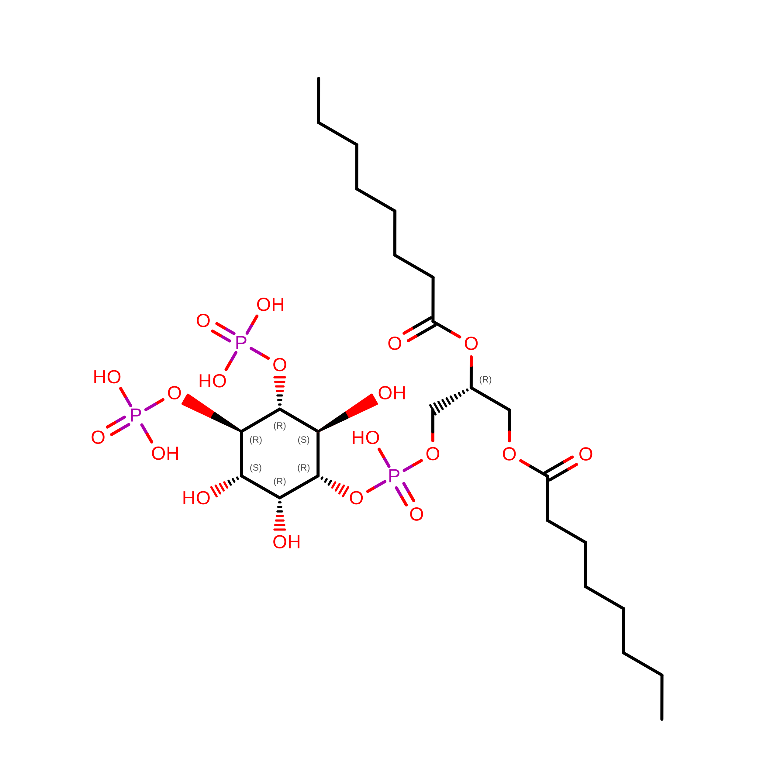
Different inositol phospholipids (PIPs) distribute to distinct subcellular organelles, creating an addressing system that dictates the sites of action of PIP-binding proteins, including components of the Endosomal Sorting Complexes Required for Transport (ESCRT). The ESCRT machinery is recruited to remodel many different cellular membranes through combinatorial binding interactions made by the early-acting ESCRT-I and ESCRT-II complexes with PIPs, ubiquitin modifications, and membrane-specific adaptors. Membrane remodeling, constriction, and fission are then mediated by membrane-associated filaments formed by subunits of the late-acting ESCRT-III complexes, together with their associated VPS4 AAA ATPases. Here, we describe two different classes of helical ESCRT-III filaments that can surround and tubulate membranes containing PIP 2 lipids. Cryo-EM reconstructions revealed that protofilaments comprising closed IST1 subunits formed 8-stranded nanotubes that encase membrane monolayers. The nanotube coordinates exposed PI(4,5)P 2 or PI(3,5)P 2 headgroups within a basic pocket formed at the junction of three IST1 subunits, and our structures reveal how the pocket can accommodate either PIP 2 isomer with minimal adjustment. In contrast, protofilaments comprising open CHMP1A subunits formed one start helices that encase membrane bilayers and bind exposed PI(4,5)P 2 headgroups across a basic surface that spans adjacent subunits of the CHMP1A protofilament. These two different structures extend the known plasticity of ESCRT-III polymers, reveal how PIP 2 lipids can promote ESCRT-III filament assembly and membrane remodeling, and define the molecular contacts that underlie specific ESCRT-III/PIP 2 interactions.
- Department of Biochemistry, University of Utah Spencer Fox Eccles School of Medicine, Salt Lake City, UT 84112.
Organizational Affiliation: