Find PDB structures and Computed Structure Models (CSM) by combining queries from tools in this suite: Attribute Search, Sequence Similarity, Sequence Motif, 3D Similarity, and 3D Motif with 'AND' logic.
Funding Organization(s): National Institutes of Health/National Institute of General Medical Sciences (NIH/NIGMS), Department of Energy (DOE, United States)
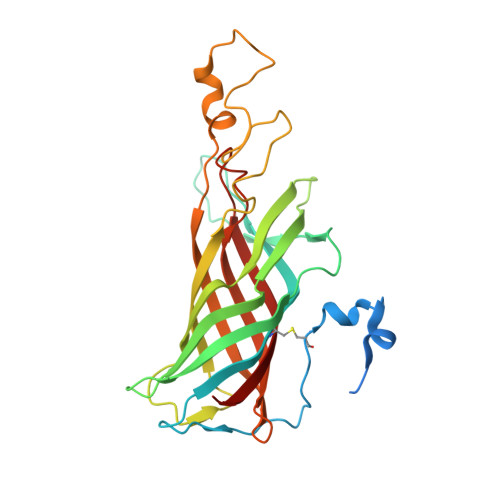
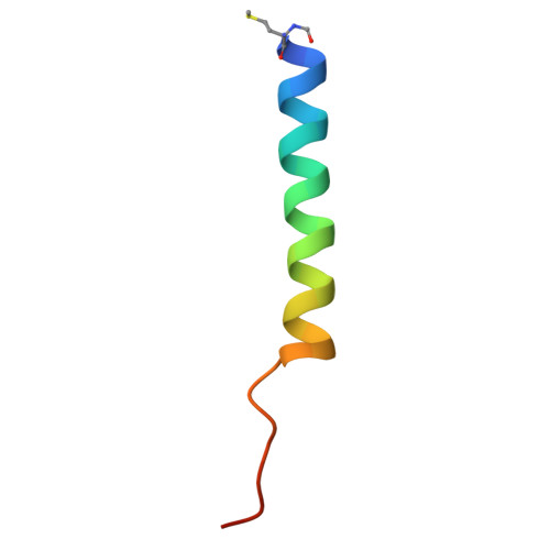
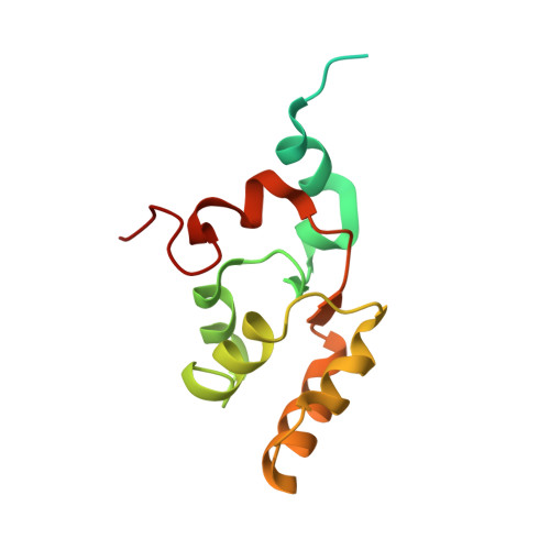
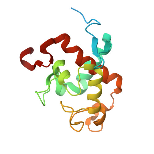
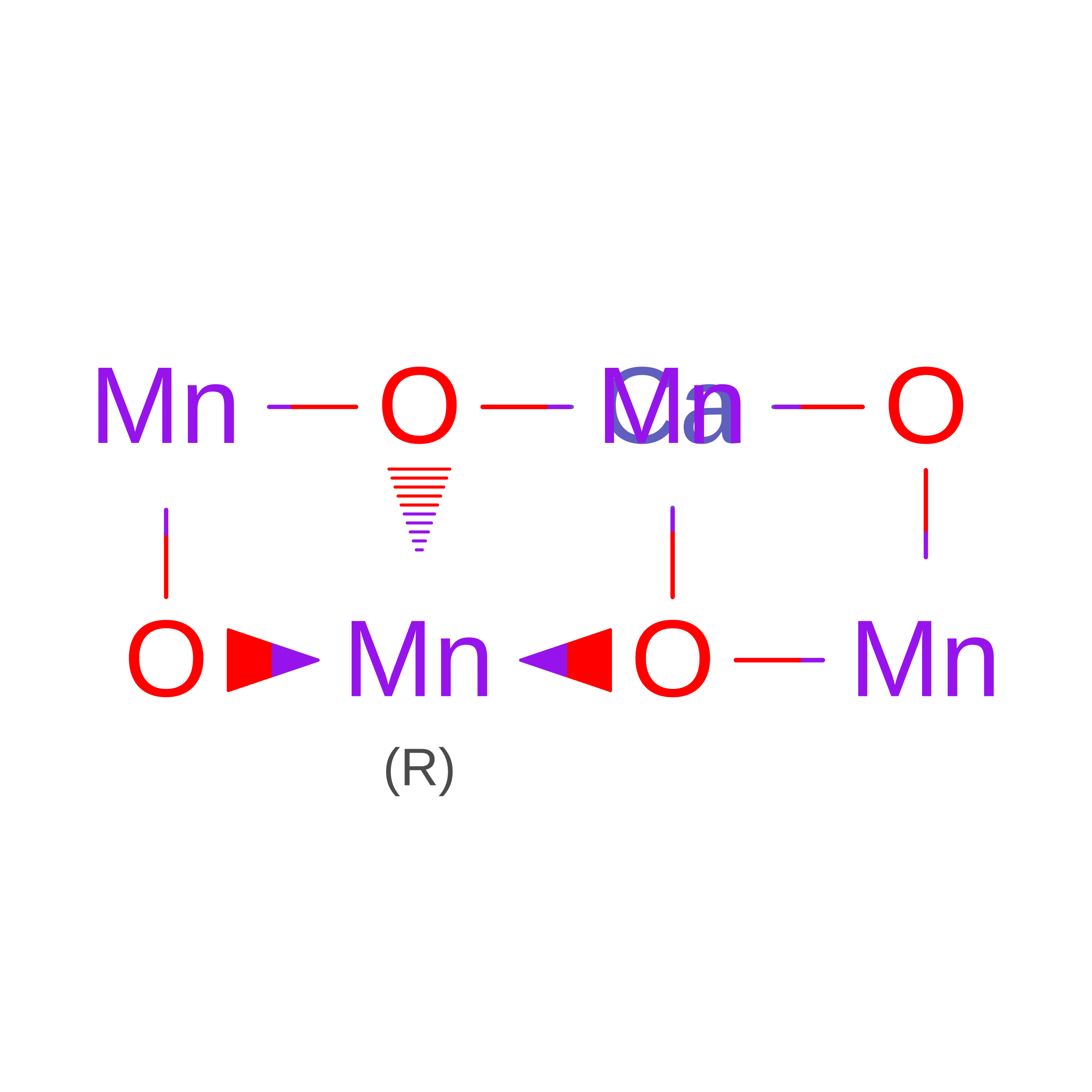
Water oxidation in photosynthesis occurs through a four-electron redox reaction that is catalyzed by the oxygen-evolving complex (OEC) in Photosystem II (PS II), that consists of the Mn 4 CaO 5 cluster. Among the four stable intermediates in the water oxidation reaction, it has been reported that the paramagnetic S 2 state with the formal oxidation of Mn(III)Mn(IV) 3 has multiple spin states, that include high spin (HS, 5/2, g≥4.1) and low spin (LS, 1/2, g = 2) forms. Previous computational studies proposed that these two forms differ in their structure and the high spin form may represent an intermediate form of the OEC during the S 2 to S 3 transition, the step in which the first substrate water binds to the last open coordination site of the cluster. In the current study, we examined the pH-induced HS-S 2 state of T. vestitus PS II with MnK edge extended X-ray absorption fine structure (EXAFS) and X-ray absorption near edge spectroscopy (XANES). Small, but notable differences were observed in the EXAFS and XANES between the HS and LS S 2 states. EXAFS data and the fits indicate that the basic geometry of the cluster remains the same between these two states. Therefore, we hypothesize that the difference between the HS and LS S 2 state may be due to chemical differences such as different protonation states of the oxo and/or water ligands, or its surroundings. We also examined the detailed oxygen positions of the S 2 state in the XFEL-based room temperature crystal structures, and confirmed that at neutral pH at room temperature, the structure is consistent with that of the LS-S 2 model with open cubane-like geometry.
Organizational Affiliation:
Molecular Biophysics and Integrated Bioimaging Division, Lawrence Berkeley National Laboratory, 1 Cyclotron Road, Berkeley, CA, 94720, USA.
Molecular Biophysics and Integrated Bioimaging Division, Lawrence Berkeley National Laboratory, 1 Cyclotron Road, Berkeley, CA, 94720, USA. Electronic address: vkyachandra@lbl.gov.
Molecular Biophysics and Integrated Bioimaging Division, Lawrence Berkeley National Laboratory, 1 Cyclotron Road, Berkeley, CA, 94720, USA. Electronic address: jyano@lbl.gov.

 Download Mendeley