11FS | pdb_000011fs

Effector complex from type IV-C CRISPR-Cas system


Experimental Data Snapshot

  • Method: ELECTRON MICROSCOPY
  • Resolution: 3.20 Å
  • Aggregation State: PARTICLE 
  • Reconstruction Method: SINGLE PARTICLE 

wwPDB Validation 3D Report Full Report

Validation slider image for 11FS

This is version 1.1 of the entry. See complete history. 

Literature

Type IV-C CRISPR-Cas effector complexes recognize double-stranded DNA and switch on collateral cleavage of ssDNA and RNA.

Pittman, C.C., Xu, C., Catchpole, R.J., Garrett, S., Fuchs, R., Chu, X., Makarova, K.S., Koonin, E.V., Zhao, P., Wells, L., Graveley, B.R., Ke, A., Terns, M.P.

(2026) Cell Rep 45: 117939-117939

  • DOI: https://doi.org/10.1016/j.celrep.2026.117939
  • Primary Citation Related Structures: 
    11FS, 11HC, 11HD

  • PubMed Abstract: 

    Type IV-C CRISPR-Cas systems remain enigmatic compared to other class 1 systems. Here, we expand the type IV-C catalog, identifying two phylogenetically distinct clades primarily found in archaea (IV-C1) or bacteria (IV-C2), distinguishable by the Cas10IVc subunit architecture. We functionally and structurally characterize type IV-C1 systems from Thermococcus onnurineus (Ton) and Pyrococcus abyssi (Pab). Type IV-C complexes assemble with crRNAs derived from distinct CRISPR arrays and recognize a 5'-GGG-3' protospacer adjacent motif (PAM) to bind double-stranded DNA targets. Target recognition activates the HD domain of Cas10IVc, triggering metal-dependent collateral cleavage of single-stranded DNA and RNA. This behavior is explained by allosteric alignment of the HD active site, triggered by PAM-dependent R-loop formation, as revealed by cryo-EM. Together, our findings suggest that type IV-C systems provide immunity via non-specific cleavage of nucleic acids generated during mobile genetic element replication or transcription.


  • Organizational Affiliation: 
    • Department of Microbiology, University of Georgia, Athens, GA 30605, USA.

Macromolecule Content 

  • Total Structure Weight: 478.62 kDa 
  • Atom Count: 30,547 
  • Modeled Residue Count: 3,694 
  • Deposited Residue Count: 4,072 
  • Unique protein chains: 4
  • Unique nucleic acid chains: 3

Macromolecules


Find similar proteins by:|  3D Structure
Entity ID: 1
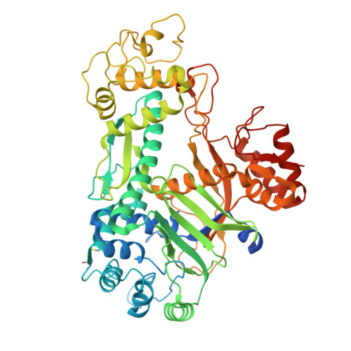
MoleculeChains  Sequence LengthOrganismDetailsImage
Cas10IVc608Pyrococcus abyssiMutation(s): 0 
Entity Groups
Sequence Clusters30% Identity50% Identity70% Identity90% Identity95% Identity100% Identity
Sequence Annotations
Expand
Reference Sequence
Find similar proteins by:|  3D Structure
Entity ID: 2
MoleculeChains  Sequence LengthOrganismDetailsImage
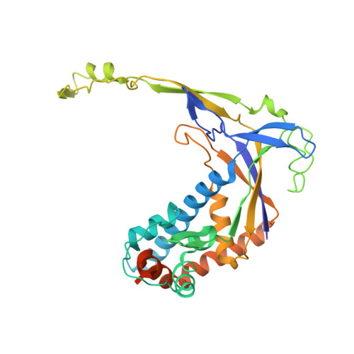
Cas7c330Pyrococcus abyssiMutation(s): 0 
Entity Groups
Sequence Clusters30% Identity50% Identity70% Identity90% Identity95% Identity100% Identity
Sequence Annotations
Expand
Reference Sequence
Find similar proteins by:|  3D Structure
Entity ID: 3
MoleculeChains  Sequence LengthOrganismDetailsImage
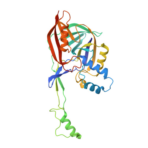
Cas5cC [auth D]306Pyrococcus abyssiMutation(s): 0 
Entity Groups
Sequence Clusters30% Identity50% Identity70% Identity90% Identity95% Identity100% Identity
Sequence Annotations
Expand
Reference Sequence
Find similar proteins by:|  3D Structure
Entity ID: 7
MoleculeChains  Sequence LengthOrganismDetailsImage
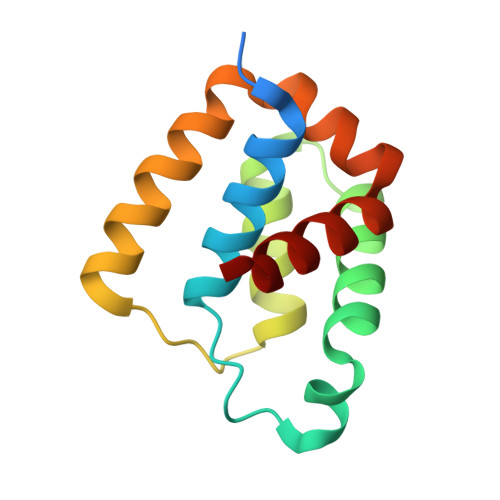
Cas11c124Pyrococcus abyssiMutation(s): 0 
Entity Groups
Sequence Clusters30% Identity50% Identity70% Identity90% Identity95% Identity100% Identity
Sequence Annotations
Expand
Reference Sequence
Find similar nucleic acids by:  Sequence
Entity ID: 4
MoleculeChains LengthOrganismImage
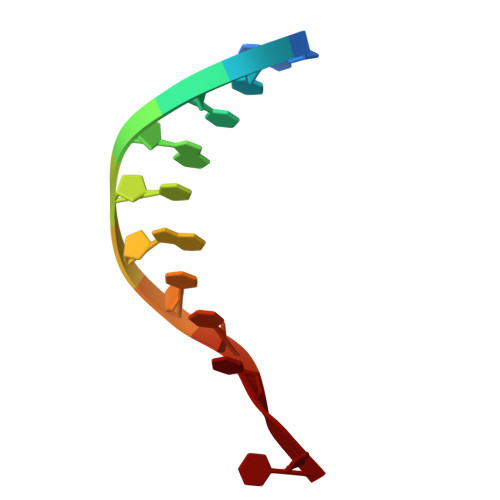
Non-Target Strand DNAD [auth E]10Pyrococcus abyssi
Sequence Annotations
Expand
Reference Sequence
Find similar nucleic acids by:  (by identity cutoff) 
Entity ID: 5
MoleculeChains LengthOrganismImage
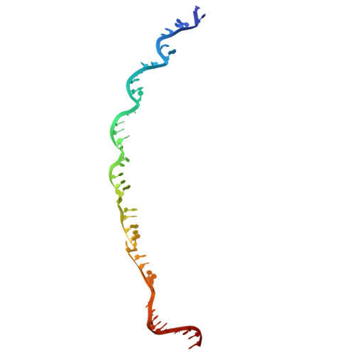
Target Strand DNAE [auth F]47Pyrococcus abyssi
Sequence Annotations
Expand
Reference Sequence
Find similar nucleic acids by:  (by identity cutoff) 
Entity ID: 6
MoleculeChains LengthOrganismImage
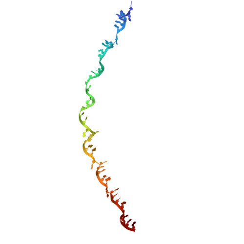
crRNA (47-MER)F [auth G]47Pyrococcus abyssi
Sequence Annotations
Expand
Reference Sequence

Experimental Data & Validation

Experimental Data

  • Method: ELECTRON MICROSCOPY
  • Resolution: 3.20 Å
  • Aggregation State: PARTICLE 
  • Reconstruction Method: SINGLE PARTICLE 
EM Software:
TaskSoftware PackageVersion
MODEL REFINEMENTPHENIX1.21.2_5419
RECONSTRUCTIONcryoSPARC

Structure Validation

View Full Validation Report


Entry History 

& Funding Information

Deposition Data


Funding OrganizationLocationGrant Number
National Institutes of Health/National Institute of General Medical Sciences (NIH/NIGMS)United StatesR35118174

Revision History  (Full details and data files)

  • Version 1.0: 2026-08-26
    Type: Initial release
  • Version 1.1: 2026-09-16
    Changes: Data collection, Database references