10SF | pdb_000010sf

The CryoEM structure of T10 type1 nanofiber


Experimental Data Snapshot

  • Method: ELECTRON MICROSCOPY
  • Resolution: 3.08 Å
  • Aggregation State: FILAMENT 
  • Reconstruction Method: HELICAL 

wwPDB Validation 3D Report Full Report

Validation slider image for 10SF

This is version 1.0 of the entry. See complete history. 

Literature

Tailoring Avidity through Morphology: Structure-Avidity Relationship in CD38-Binding Nanofiber Radiotracers.

Godbe, J.M., Zhang, H., Sharma, A.K., Ernst, K.N., Jing, Z., Dyer, M.R., Prior, J.L., Teubner, E., Manion, B., Tang, R., Yang, Y., Shokeen, M.

(2026) ACS Appl Bio Mater 9: 4242-4257

  • DOI: https://doi.org/10.1021/acsabm.6c00348
  • Primary Citation Related Structures: 
    10SD, 10SE, 10SF, 10SG, 10SH

  • PubMed Abstract: 

    The lack of targeted molecular imaging agents for multiple myeloma (MM) hinders precise disease characterization and theranostic development. We address this by engineering a tunable platform of self-assembled peptide nanofibers that target CD38, a key antigen in MM. Simple variation of a conjugated lipid tail length (C4-C12) dictates the supramolecular architecture, as revealed by high-resolution cryo-EM. This structural control directly modulates biological function: avidity for CD38 increases monotonically with tail length, culminating in T12 nanofibers with sub-nanomolar affinity. This optimized morphology also enables unique pH-responsive di-tyrosine cross-linking and, critically, facilitates polyvalent cell-surface engagement that outcompetes high-affinity monomers in vitro. The nanofibers are efficiently radiolabeled with 64 Cu, exhibit exceptional serum stability, and show no toxicity at doses 20-fold above projected imaging use. By establishing lipid tail length as a simple, powerful handle for controlling nanofiber structure, avidity, and function, we present a robust, translatable platform for advancing targeted imaging and therapy in CD38-positive malignancies.


  • Organizational Affiliation: 
    • Edward Mallinckrodt Institute of Radiology, Washington University School of Medicine, St. Louis, Missouri 63110, United States.

Macromolecule Content 

  • Total Structure Weight: 22.37 kDa 
  • Atom Count: 1,566 
  • Modeled Residue Count: 198 
  • Deposited Residue Count: 198 
  • Unique protein chains: 1

Macromolecules

Find similar proteins by:  Sequence   |   3D Structure  
Entity ID: 1
MoleculeChains  Sequence LengthOrganismDetailsImage
Type10 type 1 nanofiber11synthetic constructMutation(s): 0 
Entity Groups
Sequence Clusters30% Identity50% Identity70% Identity90% Identity95% Identity100% Identity
Sequence Annotations
Expand
Reference Sequence

Small Molecules

Ligands 1 Unique
IDChains Name / Formula / InChI Key2D Diagram3D Interactions
DKA

Query on DKA


Download:Ideal Coordinates CCD File
AA [auth I]
BA [auth J]
CA [auth K]
DA [auth L]
EA [auth M]
AA [auth I],
BA [auth J],
CA [auth K],
DA [auth L],
EA [auth M],
FA [auth N],
GA [auth O],
HA [auth P],
IA [auth Q],
JA [auth R],
S [auth E],
T [auth A],
U [auth B],
V [auth C],
W [auth D],
X [auth F],
Y [auth G],
Z [auth H]
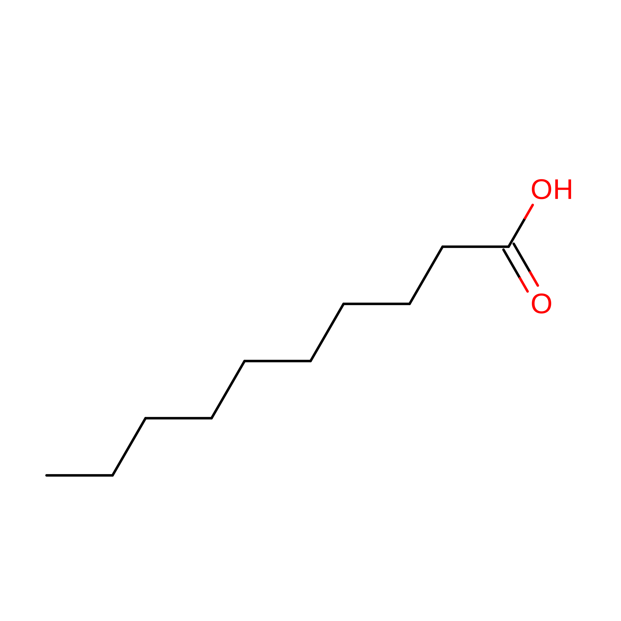
DECANOIC ACID
C10 H20 O2
GHVNFZFCNZKVNT-UHFFFAOYSA-N

Experimental Data & Validation

Experimental Data

  • Method: ELECTRON MICROSCOPY
  • Resolution: 3.08 Å
  • Aggregation State: FILAMENT 
  • Reconstruction Method: HELICAL 
EM Software:
TaskSoftware PackageVersion
MODEL REFINEMENTREFMAC5.8.0430
RECONSTRUCTIONServalcat

Structure Validation

View Full Validation Report


Entry History 

& Funding Information

Deposition Data


Funding OrganizationLocationGrant Number
Other private--

Revision History  (Full details and data files)

  • Version 1.0: 2026-07-01
    Type: Initial release