8UK3

The rotavirus VP5*/VP8* conformational transition permeabilizes membranes to Ca2+ (class 6 reconstruction)


Experimental Data Snapshot

  • Method: ELECTRON MICROSCOPY
  • Resolution: 8.00 Å
  • Aggregation State: PARTICLE 
  • Reconstruction Method: SINGLE PARTICLE 

wwPDB Validation   3D Report Full Report


This is version 1.1 of the entry. See complete history


Literature

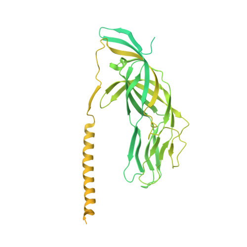
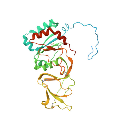
The rotavirus VP5*/VP8* conformational transition permeabilizes membranes to Ca2.

de Sautu, M.Herrmann, T.Scanavachi, G.Jenni, S.Harrison, S.C.

(2024) PLoS Pathog 20: e1011750-e1011750

  • DOI: https://doi.org/10.1371/journal.ppat.1011750
  • Primary Citation of Related Structures:  
    8UK2, 8UK3

  • PubMed Abstract: 

    Rotaviruses infect cells by delivering into the cytosol a transcriptionally active inner capsid particle (a "double-layer particle": DLP). Delivery is the function of a third, outer layer, which drives uptake from the cell surface into small vesicles from which the DLPs escape. In published work, we followed stages of rhesus rotavirus (RRV) entry by live-cell imaging and correlated them with structures from cryogenic electron microscopy and tomography (cryo-EM and cryo-ET). The virus appears to wrap itself in membrane, leading to complete engulfment and loss of Ca2+ from the vesicle produced by the wrapping. One of the outer-layer proteins, VP7, is a Ca2+-stabilized trimer; loss of Ca2+ releases both VP7 and the other outer-layer protein, VP4, from the particle. VP4, activated by cleavage into VP8* and VP5*, is a trimer that undergoes a large-scale conformational rearrangement, reminiscent of the transition that viral fusion proteins undergo to penetrate a membrane. The rearrangement of VP5* thrusts a 250-residue, C-terminal segment of each of the three subunits outward, while allowing the protein to remain attached to the virus particle and to the cell being infected. We proposed that this segment inserts into the membrane of the target cell, enabling Ca2+ to cross. In the work reported here, we show the validity of key aspects of this proposed sequence. By cryo-EM studies of liposome-attached virions ("triple-layer particles": TLPs) and single-particle fluorescence imaging of liposome-attached TLPs, we confirm insertion of the VP4 C-terminal segment into the membrane and ensuing generation of a Ca2+ "leak". The results allow us to formulate a molecular description of early events in entry. We also discuss our observations in the context of other work on double-strand RNA virus entry.


  • Organizational Affiliation

    Department of Biological Chemistry and Molecular Pharmacology, Harvard Medical School, Boston, Massachusetts, United States of America.


Macromolecules
Find similar proteins by:  (by identity cutoff)  |  3D Structure
Entity ID: 1
MoleculeChains Sequence LengthOrganismDetailsImage
Outer capsid protein VP4A [auth 1],
C [auth 2],
E [auth 3]
776Simian rotavirus A strain RRVMutation(s): 0 
UniProt
Find proteins for G0YZG6 (Rotavirus A (strain RVA/Monkey/United States/RRV/1975/G3P5B[3]))
Explore G0YZG6 
Go to UniProtKB:  G0YZG6
Entity Groups  
Sequence Clusters30% Identity50% Identity70% Identity90% Identity95% Identity100% Identity
UniProt GroupG0YZG6
Sequence Annotations
Expand
  • Reference Sequence
Find similar proteins by:  (by identity cutoff)  |  3D Structure
Entity ID: 2
MoleculeChains Sequence LengthOrganismDetailsImage
Outer capsid glycoprotein VP7326Simian rotavirus A strain RRVMutation(s): 0 
UniProt
Find proteins for P12476 (Rotavirus A (strain RVA/Monkey/United States/RRV/1975/G3P5B[3]))
Explore P12476 
Go to UniProtKB:  P12476
Entity Groups  
Sequence Clusters30% Identity50% Identity70% Identity90% Identity95% Identity100% Identity
UniProt GroupP12476
Sequence Annotations
Expand
  • Reference Sequence
Small Molecules
Ligands 2 Unique
IDChains Name / Formula / InChI Key2D Diagram3D Interactions
NAG
Query on NAG

Download Ideal Coordinates CCD File 
BB [auth n]
DA [auth h]
FC [auth c]
HB [auth o]
JA [auth i]
BB [auth n],
DA [auth h],
FC [auth c],
HB [auth o],
JA [auth i],
JB [auth p],
KC [auth d],
LC [auth e],
MA [auth j],
NB [auth q],
PA [auth k],
TB [auth r],
V [auth f],
VA [auth l],
VB [auth a],
XA [auth m],
Z [auth g],
ZB [auth b]
2-acetamido-2-deoxy-beta-D-glucopyranose
C8 H15 N O6
OVRNDRQMDRJTHS-FMDGEEDCSA-N
CA
Query on CA

Download Ideal Coordinates CCD File 
AA [auth g]
AB [auth m]
AC [auth b]
BA [auth g]
BC [auth b]
AA [auth g],
AB [auth m],
AC [auth b],
BA [auth g],
BC [auth b],
CA [auth g],
CB [auth n],
CC [auth b],
DB [auth n],
DC [auth c],
EA [auth h],
EB [auth n],
EC [auth c],
FA [auth h],
FB [auth o],
GA [auth h],
GB [auth o],
GC [auth c],
HA [auth i],
HC [auth d],
IA [auth i],
IB [auth o],
IC [auth d],
JC [auth d],
KA [auth i],
KB [auth p],
LA [auth j],
LB [auth p],
MB [auth p],
MC [auth e],
NA [auth j],
NC [auth e],
OA [auth j],
OB [auth q],
OC [auth e],
PB [auth q],
QA [auth k],
QB [auth q],
RA [auth k],
RB [auth r],
SA [auth k],
SB [auth r],
TA [auth l],
UA [auth l],
UB [auth r],
W [auth f],
WA [auth l],
WB [auth a],
X [auth f],
XB [auth a],
Y [auth f],
YA [auth m],
YB [auth a],
ZA [auth m]
CALCIUM ION
Ca
BHPQYMZQTOCNFJ-UHFFFAOYSA-N
Experimental Data & Validation

Experimental Data

  • Method: ELECTRON MICROSCOPY
  • Resolution: 8.00 Å
  • Aggregation State: PARTICLE 
  • Reconstruction Method: SINGLE PARTICLE 

Structure Validation

View Full Validation Report


Entry History & Funding Information

Deposition Data


Funding OrganizationLocationGrant Number
National Institutes of Health/National Cancer Institute (NIH/NCI)United StatesCA13202

Revision History  (Full details and data files)

  • Version 1.0: 2024-03-27
    Type: Initial release
  • Version 1.1: 2024-04-17
    Changes: Database references