4U9U | pdb_00004u9u

Crystal structure of NqrF FAD-binding domain from Vibrio cholerae


Experimental Data Snapshot

  • Method: X-RAY DIFFRACTION
  • Resolution: 1.55 Å
  • R-Value Free: 
    0.196 (Depositor), 0.190 (DCC) 
  • R-Value Work: 
    0.162 (Depositor), 0.160 (DCC) 
  • R-Value Observed: 
    0.164 (Depositor) 

wwPDB Validation   3D Report Full Report


Ligand Structure Quality Assessment 

Created with Raphaël 2.3.0Worse 01 BetterLigand structure goodness of fit to experimental dataBest fitted FADClick on this verticalbar to view details

This is version 1.2 of the entry. See complete history


Literature

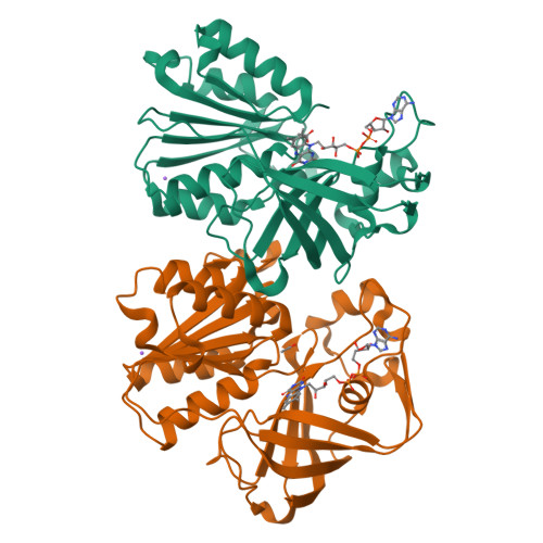
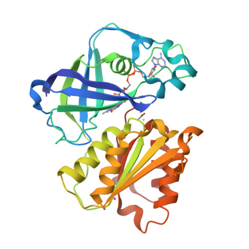
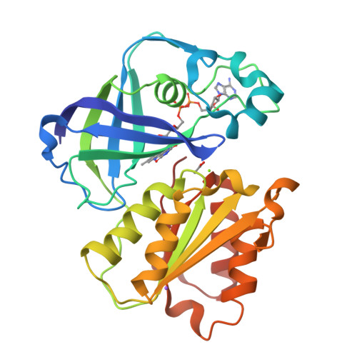
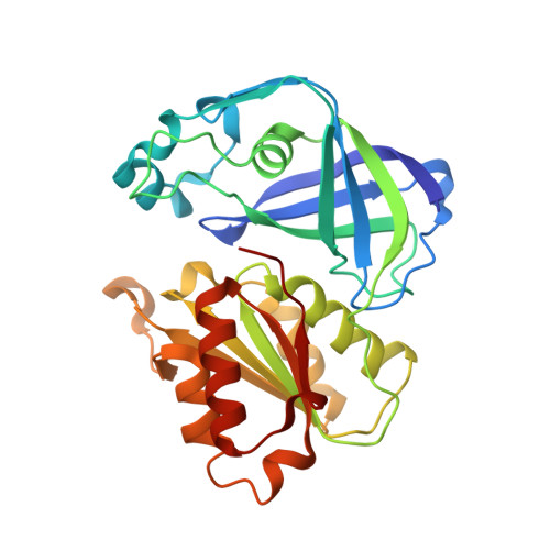
Structure of the V. cholerae Na+-pumping NADH:quinone oxidoreductase.

Steuber, J.Vohl, G.Casutt, M.S.Vorburger, T.Diederichs, K.Fritz, G.

(2014) Nature 516: 62-67

  • DOI: https://doi.org/10.1038/nature14003
  • Primary Citation of Related Structures:  
    4U9O, 4U9Q, 4U9S, 4U9U, 4UAJ

  • PubMed Abstract: 

    NADH oxidation in the respiratory chain is coupled to ion translocation across the membrane to build up an electrochemical gradient. The sodium-translocating NADH:quinone oxidoreductase (Na(+)-NQR), a membrane protein complex widespread among pathogenic bacteria, consists of six subunits, NqrA, B, C, D, E and F. To our knowledge, no structural information on the Na(+)-NQR complex has been available until now. Here we present the crystal structure of the Na(+)-NQR complex at 3.5 Å resolution. The arrangement of cofactors both at the cytoplasmic and the periplasmic side of the complex, together with a hitherto unknown iron centre in the midst of the membrane-embedded part, reveals an electron transfer pathway from the NADH-oxidizing cytoplasmic NqrF subunit across the membrane to the periplasmic NqrC, and back to the quinone reduction site on NqrA located in the cytoplasm. A sodium channel was localized in subunit NqrB, which represents the largest membrane subunit of the Na(+)-NQR and is structurally related to urea and ammonia transporters. On the basis of the structure we propose a mechanism of redox-driven Na(+) translocation where the change in redox state of the flavin mononucleotide cofactor in NqrB triggers the transport of Na(+) through the observed channel.


  • Organizational Affiliation

    Department of Microbiology, Garbenstrasse 30, University of Hohenheim, 70599 Stuttgart, Germany.


Macromolecules
Find similar proteins by:  (by identity cutoff)  |  3D Structure
Entity ID: 1
MoleculeChains Sequence LengthOrganismDetailsImage
Na(+)-translocating NADH-quinone reductase subunit F
A, B
281Vibrio choleraeMutation(s): 0 
Gene Names: nqrFVC_2290
EC: 1.6.5 (PDB Primary Data), 7.2.1.1 (UniProt)
Membrane Entity: Yes 
UniProt
Find proteins for Q9X4Q8 (Vibrio cholerae serotype O1 (strain ATCC 39315 / El Tor Inaba N16961))
Explore Q9X4Q8 
Go to UniProtKB:  Q9X4Q8
Entity Groups  
Sequence Clusters30% Identity50% Identity70% Identity90% Identity95% Identity100% Identity
UniProt GroupQ9X4Q8
Sequence Annotations
Expand
  • Reference Sequence
Small Molecules
Ligands 4 Unique
IDChains Name / Formula / InChI Key2D Diagram3D Interactions
FAD
Query on FAD

Download Ideal Coordinates CCD File 
C [auth A],
G [auth B]
FLAVIN-ADENINE DINUCLEOTIDE
C27 H33 N9 O15 P2
VWWQXMAJTJZDQX-UYBVJOGSSA-N
ACT
Query on ACT

Download Ideal Coordinates CCD File 
D [auth A],
H [auth B]
ACETATE ION
C2 H3 O2
QTBSBXVTEAMEQO-UHFFFAOYSA-M
MG
Query on MG

Download Ideal Coordinates CCD File 
E [auth A],
I [auth B]
MAGNESIUM ION
Mg
JLVVSXFLKOJNIY-UHFFFAOYSA-N
NA
Query on NA

Download Ideal Coordinates CCD File 
F [auth A],
J [auth B]
SODIUM ION
Na
FKNQFGJONOIPTF-UHFFFAOYSA-N
Experimental Data & Validation

Experimental Data

  • Method: X-RAY DIFFRACTION
  • Resolution: 1.55 Å
  • R-Value Free:  0.196 (Depositor), 0.190 (DCC) 
  • R-Value Work:  0.162 (Depositor), 0.160 (DCC) 
  • R-Value Observed: 0.164 (Depositor) 
Space Group: P 21 21 21
Unit Cell:
Length ( Å )Angle ( ˚ )
a = 73.43α = 90
b = 89.07β = 90
c = 90.48γ = 90
Software Package:
Software NamePurpose
XDSdata reduction
SOLVEphasing
XSCALEdata scaling
PDB_EXTRACTdata extraction
Cootmodel building
PHENIXrefinement
XSCALEdata reduction

Structure Validation

View Full Validation Report


Ligand Structure Quality Assessment 

Created with Raphaël 2.3.0Worse 01 BetterLigand structure goodness of fit to experimental dataBest fitted FADClick on this verticalbar to view details

Entry History 

Deposition Data

  • Released Date: 2014-12-03 
  • Deposition Author(s): Fritz, G.

Revision History  (Full details and data files)

  • Version 1.0: 2014-12-03
    Type: Initial release
  • Version 1.1: 2014-12-17
    Changes: Database references
  • Version 1.2: 2024-05-08
    Changes: Data collection, Database references, Derived calculations