4TZC | pdb_00004tzc

Crystal Structure of Murine Cereblon in Complex with Thalidomide


Experimental Data Snapshot

  • Method: X-RAY DIFFRACTION
  • Resolution: 1.88 Å
  • R-Value Free: 
    0.220 (Depositor), 0.220 (DCC) 
  • R-Value Work: 
    0.173 (Depositor), 0.170 (DCC) 
  • R-Value Observed: 
    0.175 (Depositor) 

wwPDB Validation   3D Report Full Report


Ligand Structure Quality Assessment 

Created with Raphaël 2.3.0Worse 01 BetterLigand structure goodness of fit to experimental dataBest fitted EF2Click on this verticalbar to view details

This is version 1.3 of the entry. See complete history


Literature

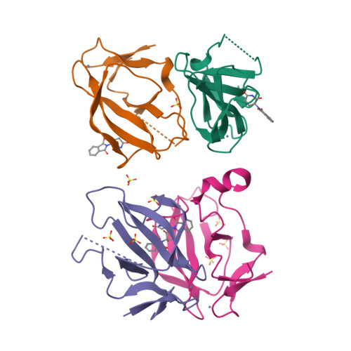
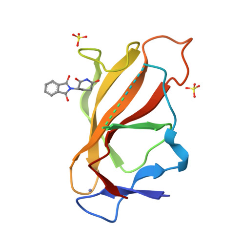
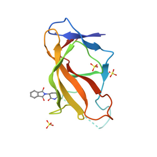
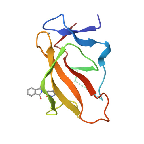
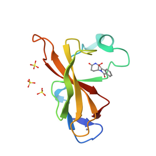
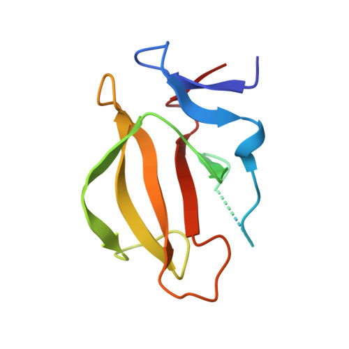
Structural Basis for Responsiveness to Thalidomide-Analog Drugs Defined by the Crystal Structure of the Human Cereblon:DDB1:Lenalidomide Complex

Chamberlain, P.P.Lopez-Girona, A.Miller, K.Carmel, G.Pagarigan, B.Leon, B.Rychak, E.Corral, L.Ren, Y.Wang, M.Riley, M.Delker, S.Ito, T.Hideki, A.Mori, T.Hirano, Y.Handa, H.Hakoshima, T.Daniel, T.O.Cathers, B.E.

To be published.

Macromolecules
Find similar proteins by:  (by identity cutoff)  |  3D Structure
Entity ID: 1
MoleculeChains Sequence LengthOrganismDetailsImage
Protein cereblonA [auth C],
B [auth A],
C [auth B],
D
108Mus musculusMutation(s): 0 
Gene Names: Crbn
UniProt & NIH Common Fund Data Resources
Find proteins for Q8C7D2 (Mus musculus)
Explore Q8C7D2 
Go to UniProtKB:  Q8C7D2
IMPC:  MGI:1913277
Entity Groups  
Sequence Clusters30% Identity50% Identity70% Identity90% Identity95% Identity100% Identity
UniProt GroupQ8C7D2
Sequence Annotations
Expand
  • Reference Sequence
Small Molecules
Ligands 3 Unique
IDChains Name / Formula / InChI Key2D Diagram3D Interactions
EF2
Query on EF2

Download Ideal Coordinates CCD File 
F [auth C],
J [auth A],
O [auth B],
T [auth D]
S-Thalidomide
C13 H10 N2 O4
UEJJHQNACJXSKW-VIFPVBQESA-N
SO4
Query on SO4

Download Ideal Coordinates CCD File 
H [auth A]
I [auth A]
L [auth B]
M [auth B]
N [auth B]
SULFATE ION
O4 S
QAOWNCQODCNURD-UHFFFAOYSA-L
ZN
Query on ZN

Download Ideal Coordinates CCD File 
E [auth C],
G [auth A],
K [auth B],
P [auth D]
ZINC ION
Zn
PTFCDOFLOPIGGS-UHFFFAOYSA-N
Binding Affinity Annotations 
IDSourceBinding Affinity
EF2 BindingDB:  4TZC Ki: min: 1800, max: 2.27e+4 (nM) from 5 assay(s)
IC50: min: 268, max: 2.99e+4 (nM) from 7 assay(s)
Experimental Data & Validation

Experimental Data

  • Method: X-RAY DIFFRACTION
  • Resolution: 1.88 Å
  • R-Value Free:  0.220 (Depositor), 0.220 (DCC) 
  • R-Value Work:  0.173 (Depositor), 0.170 (DCC) 
  • R-Value Observed: 0.175 (Depositor) 
Space Group: I 2 3
Unit Cell:
Length ( Å )Angle ( ˚ )
a = 143.146α = 90
b = 143.146β = 90
c = 143.146γ = 90
Software Package:
Software NamePurpose
REFMACrefinement
HKL-2000data reduction
PDB_EXTRACTdata extraction
DENZOdata reduction
SCALEPACKdata scaling

Structure Validation

View Full Validation Report


Ligand Structure Quality Assessment 

Created with Raphaël 2.3.0Worse 01 BetterLigand structure goodness of fit to experimental dataBest fitted EF2Click on this verticalbar to view details

Entry History 

Deposition Data

Revision History  (Full details and data files)

  • Version 1.0: 2014-08-06
    Type: Initial release
  • Version 1.1: 2016-03-16
    Changes: Data collection
  • Version 1.2: 2017-09-27
    Changes: Derived calculations, Refinement description
  • Version 1.3: 2023-12-27
    Changes: Data collection, Database references