9YXN | pdb_00009yxn

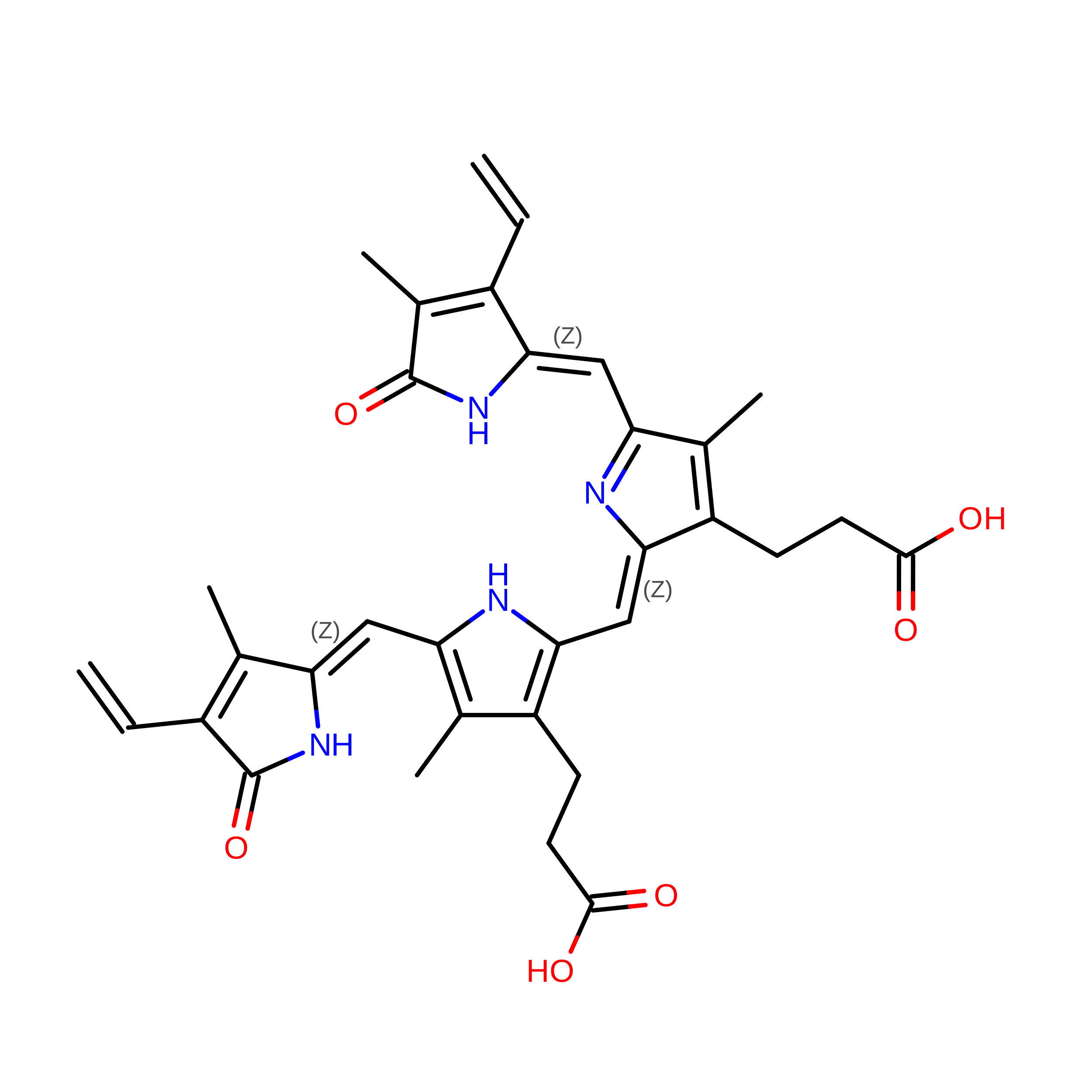
Pr-Pfr heterodimer state of Stigmatella aurantiaca bacteriophytochrome 2


Experimental Data Snapshot

  • Method: ELECTRON MICROSCOPY
  • Resolution: 5.01 Å
  • Aggregation State: CELL 
  • Reconstruction Method: SINGLE PARTICLE 

wwPDB Validation 3D Report Full Report

Validation slider image for 9YXN

This is version 1.2 of the entry. See complete history. 

Literature

A bacteriophytochrome Pr/Pfr heterodimer studied through single-particle time-resolved cryo-electron microscopy.

Karki, P., Budell, W.C., Kannachel, R., Menendez, D., Hernandez, C., Mendez, J.H., Kanou, S., Singh, M., Slavov, C., Schwander, P., Malla, T.N., Stojkovic, E.A., Schmidt, M.

(2026) Commun Chem 9

  • DOI: https://doi.org/10.1038/s42004-026-02084-6
  • Primary Citation Related Structures: 
    9YS3, 9YXN

  • PubMed Abstract: 

    Phytochromes are dimeric photoreceptors found in bacteria, fungi, and plants that reversibly interconvert between a red-absorbing Pr state and a far-red-absorbing Pfr state. In bacteria, phytochromes (BphPs) regulate diverse responses through a two-component signaling pathway comprising a C-terminal histidine kinase (HK) and a response regulator. A previous cryo-EM study of the wild-type BphP from Stigmatella aurantiaca (SaBphP2) revealed a stable Pr/Pfr heterodimer in which the two protomers adopt distinct Pr and Pfr conformations. Here, using the Spotiton technique, we captured the same heterodimer by illuminating SaBphP2 particles on the cryo-EM grid and vitrifying them 10 ms later. Comparison with the Pr/Pr homodimer reveals a 180° rotation of the HK domain, driven by an unwinding of the coiled-coil helices that connect the photosensory core to the enzymatic domain. The large-scale reorientations provide mechanistic insight into light-triggered signal transduction mediated by bacterial phytochromes.


  • Organizational Affiliation: 
    • Department of Physics and Astronomy, University of Wisconsin-Milwaukee, Milwaukee, WI, USA.

Macromolecule Content 

  • Total Structure Weight: 161.66 kDa 
  • Atom Count: 10,153 
  • Modeled Residue Count: 1,478 
  • Deposited Residue Count: 1,478 
  • Unique protein chains: 1

Macromolecules

Find similar proteins by:|  3D Structure
Entity ID: 1
MoleculeChains  Sequence LengthOrganismDetailsImage
Bacteriophytochrome
A, B
739Allostigmatella aurantiacaMutation(s): 0 
Gene Names: SAMN05444354_11920
EC: 2.7.13.3
UniProt
Find proteins for A0A1H7ZJA8 (Stigmatella aurantiaca)
Explore A0A1H7ZJA8 
Go to UniProtKB:  A0A1H7ZJA8
Entity Groups
Sequence Clusters30% Identity50% Identity70% Identity90% Identity95% Identity100% Identity
UniProt GroupA0A1H7ZJA8
Sequence Annotations
Expand
Reference Sequence

Experimental Data & Validation

Experimental Data

  • Method: ELECTRON MICROSCOPY
  • Resolution: 5.01 Å
  • Aggregation State: CELL 
  • Reconstruction Method: SINGLE PARTICLE 
EM Software:
TaskSoftware PackageVersion
MODEL REFINEMENTPHENIX1.21.2_5419
RECONSTRUCTIONcryoSPARCv4.7.1

Structure Validation

View Full Validation Report


Entry History 

& Funding Information

Deposition Data


Funding OrganizationLocationGrant Number
National Science Foundation (NSF, United States)United States--
Simons FoundationUnited States--
National Institutes of Health/National Institute of General Medical Sciences (NIH/NIGMS)United States--

Revision History  (Full details and data files)

  • Version 1.0: 2026-06-17
    Type: Initial release
  • Version 1.1: 2026-06-24
    Changes: Data collection, Database references
  • Version 1.2: 2026-09-30
    Changes: Data collection, Database references