9YS3 | pdb_00009ys3

Pr-pr homodimer state of Stigmatella aurantiaca bacteriophytochrome 2


Experimental Data Snapshot

  • Method: ELECTRON MICROSCOPY
  • Resolution: 5.85 Å
  • Aggregation State: CELL 
  • Reconstruction Method: SINGLE PARTICLE 

wwPDB Validation 3D Report Full Report

Validation slider image for 9YS3

This is version 1.1 of the entry. See complete history

Literature

A bacteriophytochrome Pr/Pfr heterodimer studied through single-particle time-resolved cryo-electron microscopy.

Karki, P.Budell, W.C.Kannachel, R.Menendez, D.Hernandez, C.Mendez, J.H.Kanou, S.Singh, M.Slavov, C.Schwander, P.Malla, T.N.Stojkovic, E.A.Schmidt, M.

(2026) Commun Chem 

  • DOI: https://doi.org/10.1038/s42004-026-02084-6
  • Primary Citation Related Structures: 
    9YS3, 9YXN

  • PubMed Abstract: 

    Phytochromes are dimeric photoreceptors found in bacteria, fungi, and plants that reversibly interconvert between a red-absorbing Pr state and a far-red-absorbing Pfr state. In bacteria, phytochromes (BphPs) regulate diverse responses through a two-component signaling pathway comprising a C-terminal histidine kinase (HK) and a response regulator. A previous cryo-EM study of the wild-type BphP from Stigmatella aurantiaca (SaBphP2) revealed a stable Pr/Pfr heterodimer in which the two protomers adopt distinct Pr and Pfr conformations. Here, using the Spotiton technique, we captured the same heterodimer by illuminating SaBphP2 particles on the cryo-EM grid and vitrifying them 10 ms later. Comparison with the Pr/Pr homodimer reveals a 180° rotation of the HK domain, driven by an unwinding of the coiled-coil helices that connect the photosensory core to the enzymatic domain. The large-scale reorientations provide mechanistic insight into light-triggered signal transduction mediated by bacterial phytochromes.


  • Organizational Affiliation
    • Department of Physics and Astronomy, University of Wisconsin-Milwaukee, Milwaukee, WI, USA.

Macromolecule Content 

  • Total Structure Weight: 161.66 kDa 
  • Atom Count: 10,166 
  • Modeled Residue Count: 1,478 
  • Deposited Residue Count: 1,478 
  • Unique protein chains: 1

Macromolecules

Find similar proteins by:|  3D Structure
Entity ID: 1
MoleculeChains  Sequence LengthOrganismDetailsImage
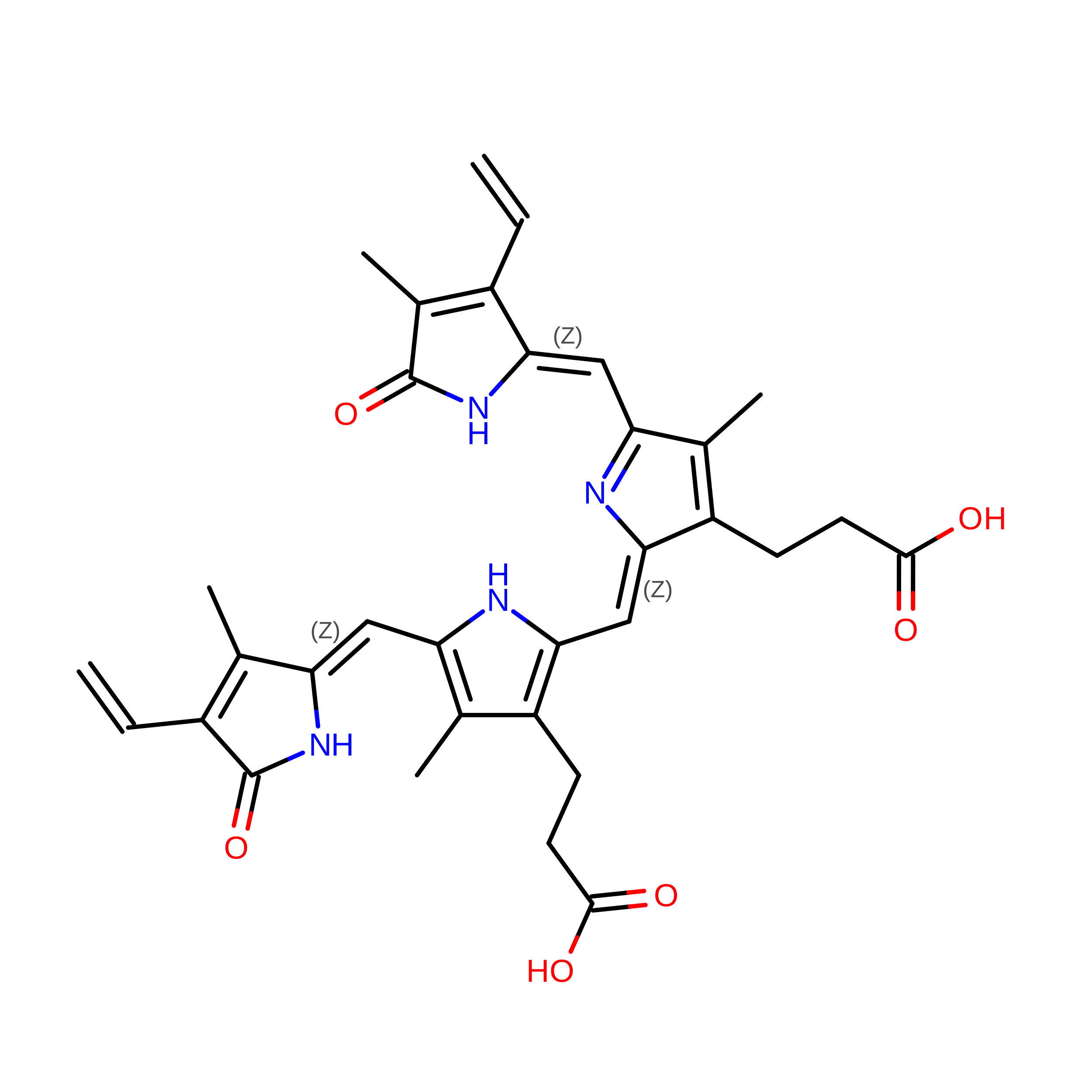
Bacteriophytochrome
A, B
739Allostigmatella aurantiacaMutation(s): 0 
Gene Names: SAMN05444354_11920
EC: 2.7.13.3
UniProt
Find proteins for A0A1H7ZJA8 (Stigmatella aurantiaca)
Explore A0A1H7ZJA8 
Go to UniProtKB:  A0A1H7ZJA8
Entity Groups
Sequence Clusters30% Identity50% Identity70% Identity90% Identity95% Identity100% Identity
UniProt GroupA0A1H7ZJA8
Sequence Annotations
Expand
Reference Sequence

Experimental Data & Validation

Experimental Data

  • Method: ELECTRON MICROSCOPY
  • Resolution: 5.85 Å
  • Aggregation State: CELL 
  • Reconstruction Method: SINGLE PARTICLE 
EM Software:
TaskSoftware PackageVersion
MODEL REFINEMENTPHENIX1.21.2_5419
RECONSTRUCTIONcryoSPARCv4.7.1

Structure Validation

View Full Validation Report


Entry History 

& Funding Information

Deposition Data


Funding OrganizationLocationGrant Number
National Science Foundation (NSF, United States)United States--
Simons FoundationUnited States--
National Institutes of Health/National Institute of General Medical Sciences (NIH/NIGMS)United States--

Revision History  (Full details and data files)

  • Version 1.0: 2026-06-17
    Type: Initial release
  • Version 1.1: 2026-06-24
    Changes: Data collection, Database references