9YPB | pdb_00009ypb

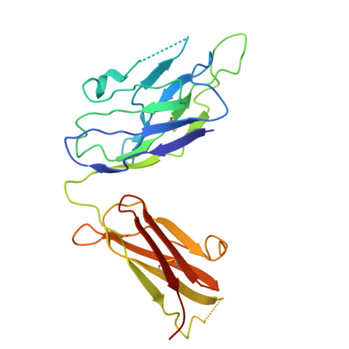
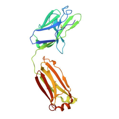
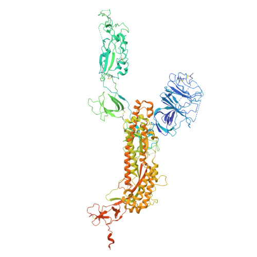
Fab-14/SARS-CoV-2 D614G spike complex, Mode IV, subgroup II conformation


Experimental Data Snapshot

  • Method: ELECTRON MICROSCOPY
  • Resolution: 3.50 Å
  • Aggregation State: PARTICLE 
  • Reconstruction Method: SINGLE PARTICLE 

wwPDB Validation 3D Report Full Report

Validation slider image for 9YPB

This is version 1.2 of the entry. See complete history. 

Literature

Neutralization of SARS-CoV-2 by IgM-14 via engagement of two distinct spike epitopes.

Wang, Y., Hu, Y., Ku, Z., Yeung, J., Zou, J., Woodson, M., Prokhorov, N.S., Knyazhanskaya, E.S., Zhao, H., Sherman, M.B., An, Z., Carroll, S.F., Shi, P.Y., Leiman, P.G., Xie, X.

(2026) PLoS Pathog 22: e1014071-e1014071

  • DOI: https://doi.org/10.1371/journal.ppat.1014071
  • Primary Citation Related Structures: 
    9YNR, 9YNX, 9YOK, 9YPB, 9YPR

  • PubMed Abstract: 

    Engineered immunoglobulin M (IgM) antibodies typically exhibit superior neutralization potency and avidity compared to their parental IgG counterparts, primarily due to multivalent binding to repeated epitopes on a targeting antigen. In this study, we characterize the neutralization breadth and mechanism of action of IgM-14, a previously reported intranasally deliverable antibody targeting SARS-CoV-2. IgM-14 demonstrates remarkably potent antiviral activity against all pre-Omicron variants but significantly reduced efficacy against Omicron BA.1, and complete loss of activity against the later subvariant JN.1. Resistance selection identified two key mutations in the receptor-binding domain (RBD), G476D and F486P, which disrupt IgM-14 binding and confer strong resistance. Cryo-electron microscopy analysis uncovered two distinct Fab-RBD interfaces: a primary interface overlapping the angiotensin-converting enzyme 2 (ACE2)-binding region, and a unique secondary interface formed only when the RBD adopts the ACE2-inaccessible "down" conformation, involving a neighboring spike protomer. Site-directed mutagenesis and structural modeling revealed a critical role of this secondary site in IgM-14-mediated neutralization. Unlike IgG-14, structural modeling suggested that IgM-14 can simultaneously engage both interfaces in diverse modes, indicating a noncanonical avidity mechanism. Collectively, these findings highlight the structural and functional uniqueness of IgM-14 and offer valuable insights into the rational design of next-generation spike-targeted antibody therapeutics with enhanced breadth and potency.


  • Organizational Affiliation: 
    • Department of Microbiology and Immunology, University of Texas Medical Branch, Galveston, Texas, United States of America.

Macromolecule Content 

  • Total Structure Weight: 528.76 kDa 
  • Atom Count: 29,858 
  • Modeled Residue Count: 3,804 
  • Deposited Residue Count: 4,760 
  • Unique protein chains: 3

Macromolecules

Find similar proteins by:|  3D Structure
Entity ID: 1
MoleculeChains  Sequence LengthOrganismDetailsImage
Fab-14 heavy chainA [auth D],
C [auth F]
235Homo sapiensMutation(s): 0 
Entity Groups
Sequence Clusters30% Identity50% Identity70% Identity90% Identity95% Identity100% Identity
Sequence Annotations
Expand
Reference Sequence
Find similar proteins by:|  3D Structure
Entity ID: 2
MoleculeChains  Sequence LengthOrganismDetailsImage
Fab-14 light chainB [auth E],
D [auth G]
213Homo sapiensMutation(s): 0 
Entity Groups
Sequence Clusters30% Identity50% Identity70% Identity90% Identity95% Identity100% Identity
Sequence Annotations
Expand
Reference Sequence
Find similar proteins by:|  3D Structure
Entity ID: 3
MoleculeChains  Sequence LengthOrganismDetailsImage
Spike glycoproteinE [auth A],
F [auth B],
G [auth C]
1,288Severe acute respiratory syndrome coronavirus 2Mutation(s): 1 
Gene Names: S, 2
UniProt
Find proteins for P0DTC2 (Severe acute respiratory syndrome coronavirus 2)
Explore P0DTC2 
Go to UniProtKB:  P0DTC2
Entity Groups
Sequence Clusters30% Identity50% Identity70% Identity90% Identity95% Identity100% Identity
UniProt GroupP0DTC2
Glycosylation
Glycosylation Sites: 6Go to GlyGen: P0DTC2-1
Sequence Annotations
Expand
Reference Sequence

Small Molecules

Ligands 1 Unique
IDChains Name / Formula / InChI Key2D Diagram3D Interactions
NAG
(Subject of Investigation/LOI)

Query on NAG


Download:Ideal Coordinates CCD File
AA [auth C]
BA [auth C]
CA [auth C]
DA [auth C]
EA [auth C]
AA [auth C],
BA [auth C],
CA [auth C],
DA [auth C],
EA [auth C],
FA [auth C],
H [auth A],
I [auth A],
J [auth A],
K [auth A],
L [auth A],
M [auth A],
N [auth A],
O [auth A],
P [auth A],
Q [auth B],
R [auth B],
S [auth B],
T [auth B],
U [auth B],
V [auth B],
W [auth B],
X [auth B],
Y [auth B],
Z [auth B]
2-acetamido-2-deoxy-beta-D-glucopyranose
C8 H15 N O6
OVRNDRQMDRJTHS-FMDGEEDCSA-N

Experimental Data & Validation

Experimental Data

  • Method: ELECTRON MICROSCOPY
  • Resolution: 3.50 Å
  • Aggregation State: PARTICLE 
  • Reconstruction Method: SINGLE PARTICLE 
EM Software:
TaskSoftware PackageVersion
MODEL REFINEMENTPHENIX1.21_5207
RECONSTRUCTIONcryoSPARC

Structure Validation

View Full Validation Report


Entry History 

& Funding Information

Deposition Data


Funding OrganizationLocationGrant Number
National Institutes of Health/National Institute of General Medical Sciences (NIH/NIGMS)United StatesR01GM139034
National Institutes of Health/National Institute of General Medical Sciences (NIH/NIGMS)United StatesR35GM158090

Revision History  (Full details and data files)

  • Version 1.0: 2026-03-18
    Type: Initial release
  • Version 1.1: 2026-03-25
    Changes: Data collection
  • Version 1.2: 2026-04-08
    Changes: Data collection, Database references