Download Mendeley
Ligand regulation and function of preformed EGFR dimers.
Zuo, Y., Schwartz, H.T., Walker, K., Han, L., Sternberg, P.W., Ferguson, K.M.(2026) Proc Natl Acad Sci U S A 123: e2602436123-e2602436123
- PubMed: 42475585 Search on PubMedSearch on PubMed Central
- DOI: https://doi.org/10.1073/pnas.2602436123
- Primary Citation Related Structures:
9YOR, 9YOS, 9YOT, 9YOU, 9YOV - PubMed Abstract:
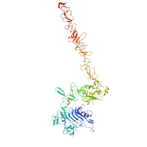
Receptor tyrosine kinases (RTKs) are key therapeutic targets in cancer, diabetes, and other diseases. With only one transmembrane α-helix-compared with seven in G-protein-coupled receptors-RTKs are thought to be activated by ligand-induced dimerization. Complicating this view, however, one of the best-studied RTKs, the insulin receptor (IR), forms allosterically regulated covalent dimers. Moreover, noncovalent "preformed" dimers have frequently been reported for the sequence-related epidermal growth factor receptor (EGFR), one of the first RTKs for which ligand-induced dimerization was described. Here, we describe a detailed structural view of a preformed EGFR dimer. Using cryo-EM, we describe how the Caenorhabditis elegans EGFR (LET-23) dimerizes without ligand. We show that preformed dimer formation modulates ligand sensitivity in vivo, but is not required for signaling itself. We also elucidate substantial ligand-induced conformational changes in LET-23 required for signaling. Our structures reveal unexpected similarities between regulation of LET-23 and the IR, suggesting that LET-23 may represent an evolutionary "missing link" between the IR and EGFR families. In the absence of ligand, intermolecular interactions within preformed receptor dimers hold the extracellular juxtamembrane regions far apart to separate the intracellular kinase domains so that they remain inactive. Ligand binding disrupts these interactions to remove the restraints on the kinase domains, which then can associate to become activated. Our analysis further suggests a unified model for the allosteric activation of preformed RTK dimers that has important implications for understanding cell-surface EGFR.
- Department of Pharmacology, Yale University School of Medicine, New Haven, CT 06520.
Organizational Affiliation: