






 Download Mendeley
Cell-Free Protein Crystallization Enables Rapid Structure Determination of Disaccharides and Trisaccharides Using Galectin-10 Crystals.
Kojima, M., Yao, X., Abe, S., Furuta, T., Hirata, K., Kobayashi, R., Suzuki, T., Ueno, T.(2025) Small Struct
Experimental Data Snapshot
Starting Model: experimental
View more details
wwPDB Validation 3D Report Full Report

(2025) Small Struct
Entity ID: 1 | |||||
|---|---|---|---|---|---|
| Molecule | Chains | Sequence Length | Organism | Details | Image |
| Galectin-10 | 142 | Homo sapiens | Mutation(s): 1 Gene Names: CLC, LGALS10, LGALS10A | 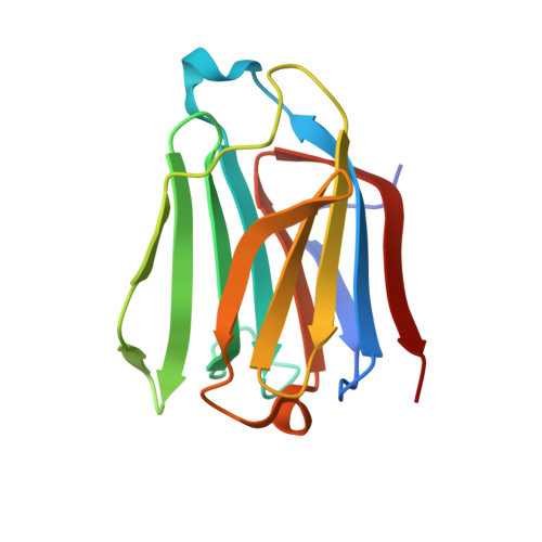 | |
UniProt & NIH Common Fund Data Resources | |||||
PHAROS: Q05315 GTEx: ENSG00000105205 | |||||
Entity Groups | |||||
| Sequence Clusters | 30% Identity50% Identity70% Identity90% Identity95% Identity100% Identity | ||||
| UniProt Group | Q05315 | ||||
Sequence AnnotationsExpand | |||||
Reference Sequence | |||||
| Ligands 1 Unique | |||||
|---|---|---|---|---|---|
| ID | Chains | Name / Formula / InChI Key | 2D Diagram | 3D Interactions | |
| EDO Download:Ideal Coordinates CCD File | C [auth A] | 1,2-ETHANEDIOL C2 H6 O2 LYCAIKOWRPUZTN-UHFFFAOYSA-N |  | ||
Entity ID: 2 | |||||
|---|---|---|---|---|---|
| ID | Chains | Name | Type/Class | 2D Diagram | 3D Interactions |
| PRD_900002 Query on PRD_900002 | B | raffinose | Oligosaccharide / Nutrient | 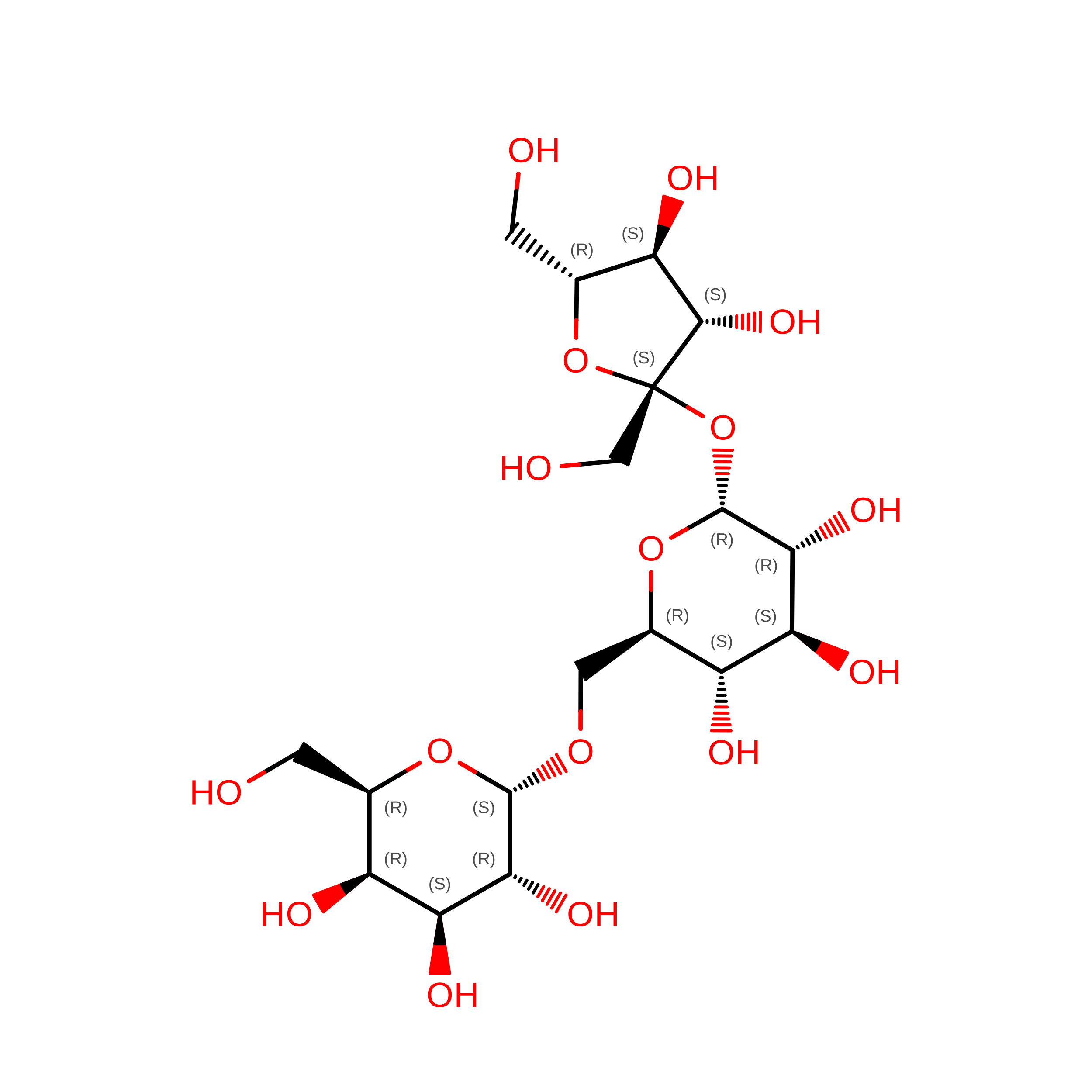 |
| Length ( Å ) | Angle ( ˚ ) |
|---|---|
| a = 49.19 | α = 90 |
| b = 49.19 | β = 90 |
| c = 261.8 | γ = 120 |
| Software Name | Purpose |
|---|---|
| PHENIX | refinement |
| Coot | model building |
| XDS | data reduction |
| XSCALE | data scaling |
| MOLREP | phasing |
| Funding Organization | Location | Grant Number |
|---|---|---|
| Japan Society for the Promotion of Science (JSPS) | Japan | -- |









