9UKM | pdb_00009ukm

Cryo-EM structure of bacteriophage P1 head


Experimental Data Snapshot

  • Method: ELECTRON MICROSCOPY
  • Resolution: 3.60 Å
  • Aggregation State: PARTICLE 
  • Reconstruction Method: SINGLE PARTICLE 

wwPDB Validation   3D Report Full Report


This is version 1.1 of the entry. See complete history


Literature

Structure of defense against restriction proteins DarA and Hdf in phage P1 reveals a new molecular mechanism during phage assembly, infection and DNA ejection.

Zheng, J.Chen, Y.Chen, S.Zhou, J.Xiao, H.Yang, F.Liu, H.

(2026) PLoS Pathog 22: e1013869-e1013869

  • DOI: https://doi.org/10.1371/journal.ppat.1013869
  • Primary Citation of Related Structures:  
    9UKM, 9UMS, 9VL4, 9VYI, 9VZ0, 9VZK

  • PubMed Abstract: 

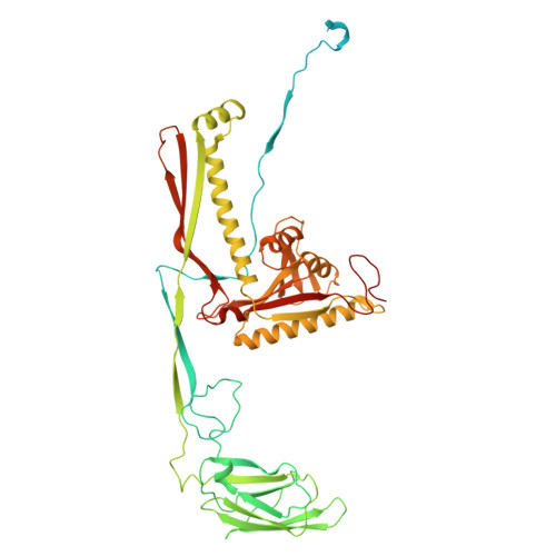
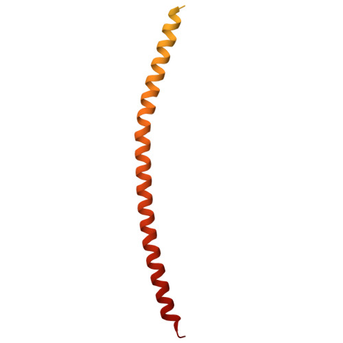
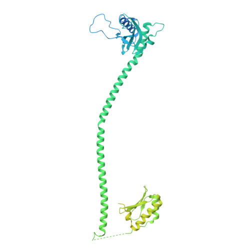
    The continuous "arms race" between bacterial antiviral defense systems and phage anti-defense strategies drives evolutionary innovation. Previous study indicated that the defense against restriction (Dar) proteins DarA and Hdf in myophage P1 are associated with the head morphogenesis. However, the structural information for these proteins was lacking, and the mechanisms by which they mediate head morphogenesis and protect phage DNA against bacterial defense systems remained poorly understood. Using cryo-electron microscopy (cryo-EM), we resolved the entire structures of extended P1 and contracted P1 with partial DNA, with the latter lacking the baseplate, as well as the head structure of contracted P1 without DNA. We identified the structural proteins for the P1, including the head, connector complex, and baseplate, which exhibited conserved properties among the majority of myophages with a simple baseplate. Notably, 55 DarA-Hdf pairs are attached to the inner surface of head at each penton-hexon junction in the extended P1 and contracted P1 with partial DNA. The DarA and Hdf together form a complex that is tightly bound to the capsid and interacts with the DNA. However, these pairs are absent in the contracted P1 without DNA. Based on our three states of P1, we hypothesis that these extensive interactions among DarA, Hdf, DNA, and head play crucial roles in mediating capsid assembly, enhancing capsid stability, and protecting phage DNA. Our results provide a structural basis for further exploration of the mechanism by which Dar proteins function during phage assembly, infection and DNA ejection. This molecular mechanism may be conserved among P1-like phages.


  • Organizational Affiliation
    • Institute of Interdisciplinary Studies, Key Laboratory for Matter Microstructure and Function of Hunan Province, Key Laboratory of Low-dimensional Quantum Structures and Quantum Control, School of Physics and Electronics, Hunan Normal University, Changsha, China.

Macromolecules
Find similar proteins by:  (by identity cutoff)  |  3D Structure
Entity ID: 1
MoleculeChains Sequence LengthOrganismDetailsImage
Gp23
A, B, C, D, E
A, B, C, D, E, F, G
568Escherichia phage P1Mutation(s): 0 
UniProt
Find proteins for A0A385AHP1 (Escherichia phage P1)
Explore A0A385AHP1 
Go to UniProtKB:  A0A385AHP1
Entity Groups  
Sequence Clusters30% Identity50% Identity70% Identity90% Identity95% Identity100% Identity
UniProt GroupA0A385AHP1
Sequence Annotations
Expand
  • Reference Sequence
Find similar proteins by:  (by identity cutoff)  |  3D Structure
Entity ID: 2
MoleculeChains Sequence LengthOrganismDetailsImage
Hdf203Escherichia phage P1Mutation(s): 0 
UniProt
Find proteins for Q71TE2 (Escherichia phage P1)
Explore Q71TE2 
Go to UniProtKB:  Q71TE2
Entity Groups  
Sequence Clusters30% Identity50% Identity70% Identity90% Identity95% Identity100% Identity
UniProt GroupQ71TE2
Sequence Annotations
Expand
  • Reference Sequence
Find similar proteins by:  (by identity cutoff)  |  3D Structure
Entity ID: 3
MoleculeChains Sequence LengthOrganismDetailsImage
Defense against restriction protein A639Escherichia phage P1Mutation(s): 0 
UniProt
Find proteins for O21970 (Escherichia phage P1)
Explore O21970 
Go to UniProtKB:  O21970
Entity Groups  
Sequence Clusters30% Identity50% Identity70% Identity90% Identity95% Identity100% Identity
UniProt GroupO21970
Sequence Annotations
Expand
  • Reference Sequence
Experimental Data & Validation

Experimental Data

  • Method: ELECTRON MICROSCOPY
  • Resolution: 3.60 Å
  • Aggregation State: PARTICLE 
  • Reconstruction Method: SINGLE PARTICLE 
EM Software:
TaskSoftware PackageVersion
MODEL REFINEMENTPHENIX1.20.1_4487

Structure Validation

View Full Validation Report


Entry History & Funding Information

Deposition Data


Funding OrganizationLocationGrant Number
National Natural Science Foundation of China (NSFC)China12034006
National Natural Science Foundation of China (NSFC)China32430020
National Natural Science Foundation of China (NSFC)China32071209

Revision History  (Full details and data files)

  • Version 1.0: 2025-10-22
    Type: Initial release
  • Version 1.1: 2026-02-04
    Changes: Data collection, Database references