9TQD | pdb_00009tqd

CD163 bound to haemoglobin


Experimental Data Snapshot

  • Method: ELECTRON MICROSCOPY
  • Resolution: 2.80 Å
  • Aggregation State: PARTICLE 
  • Reconstruction Method: SINGLE PARTICLE 

wwPDB Validation 3D Report Full Report

Validation slider image for 9TQD

This is version 1.1 of the entry. See complete history

Literature

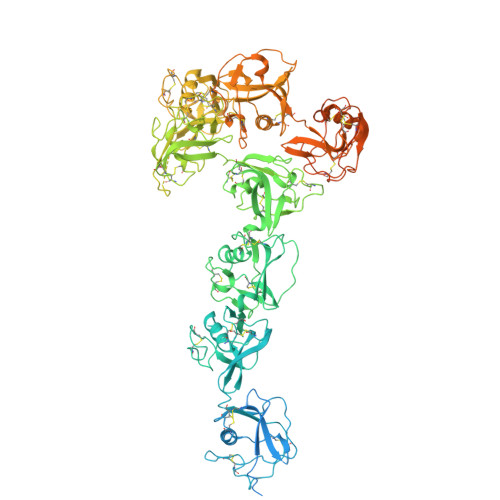
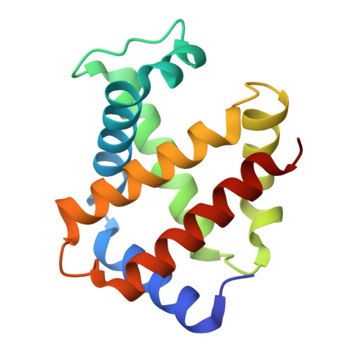
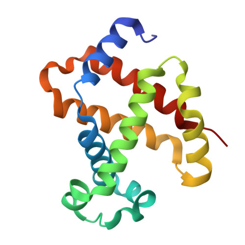
Structural basis for hemoglobin scavenging by CD163 reveals mechanism of ligand promiscuity.

Zhou, R.X.Higgins, M.K.

(2026) PLoS Biol 24: e3003788-e3003788

  • DOI: https://doi.org/10.1371/journal.pbio.3003788
  • Primary Citation Related Structures: 
    9TQD

  • PubMed Abstract: 

    The scavenger receptor CD163 detoxifies free hemoglobin released on erythrocyte lysis to prevent oxidative damage. The best understood route for hemoglobin detoxification involves the formation of haptoglobin-hemoglobin complexes that bind CD163 and are internalized into macrophages, resulting in hemoglobin degradation. However, during conditions such as sickle cell anemia or malaria, haptoglobin is depleted. CD163 can then act as a lower-affinity receptor for free hemoglobin. Previous studies revealed that CD163 forms a multimeric "base," which presents "arms" that form a binding site for haptoglobin-hemoglobin. In this study, we use cryogenic electron microscopy to reveal how human CD163 binds hemoglobin tetramers in a process that, unlike haptoglobin-hemoglobin uptake, requires a full trimeric CD163 assembly to achieve sufficient binding. We reveal how flexibility at the calcium-mediated base, combined with a hinge between receptor domains 2 and 3, allows the arms to wrap around diverse ligands. This brings together multiple small binding surfaces from different domains to form cradles for different ligands. These adaptations allow the scavenger receptor to be promiscuous, protecting us from oxidative damage caused by hemoglobin release in various pathological conditions.


  • Organizational Affiliation
    • Department of Biochemistry, University of Oxford, Oxford, United Kingdom.

Macromolecule Content 

  • Total Structure Weight: 446.52 kDa 
  • Atom Count: 24,023 
  • Modeled Residue Count: 3,110 
  • Deposited Residue Count: 4,046 
  • Unique protein chains: 3

Macromolecules

Find similar proteins by:|  3D Structure
Entity ID: 1
MoleculeChains  Sequence LengthOrganismDetailsImage
Scavenger receptor cysteine-rich type 1 protein M130
A, B, C
1,156Homo sapiensMutation(s): 0 
Gene Names: CD163M130
UniProt & NIH Common Fund Data Resources
Find proteins for Q86VB7 (Homo sapiens)
Explore Q86VB7 
Go to UniProtKB:  Q86VB7
PHAROS:  Q86VB7
GTEx:  ENSG00000177575 
Entity Groups
Sequence Clusters30% Identity50% Identity70% Identity90% Identity95% Identity100% Identity
UniProt GroupQ86VB7
Glycosylation
Glycosylation Sites: 2Go to GlyGen: Q86VB7-1
Sequence Annotations
Expand
Reference Sequence
Find similar proteins by:|  3D Structure
Entity ID: 2
MoleculeChains  Sequence LengthOrganismDetailsImage
Hemopressin
D, F
142Homo sapiensMutation(s): 0 
Gene Names: HBA1HBA2
UniProt & NIH Common Fund Data Resources
Find proteins for P69905 (Homo sapiens)
Explore P69905 
Go to UniProtKB:  P69905
Entity Groups
Sequence Clusters30% Identity50% Identity70% Identity90% Identity95% Identity100% Identity
UniProt GroupP69905
Sequence Annotations
Expand
Reference Sequence
Find similar proteins by:|  3D Structure
Entity ID: 3
MoleculeChains  Sequence LengthOrganismDetailsImage
Spinorphin
E, G
147Homo sapiensMutation(s): 0 
Gene Names: HBB
UniProt & NIH Common Fund Data Resources
Find proteins for P68871 (Homo sapiens)
Explore P68871 
Go to UniProtKB:  P68871
PHAROS:  P68871
GTEx:  ENSG00000244734 
Entity Groups
Sequence Clusters30% Identity50% Identity70% Identity90% Identity95% Identity100% Identity
UniProt GroupP68871
Sequence Annotations
Expand
Reference Sequence

Oligosaccharides

Help  
Entity ID: 4
MoleculeChains Length2D Diagram GlycosylationD Interactions
2-acetamido-2-deoxy-beta-D-glucopyranose-(1-4)-2-acetamido-2-deoxy-beta-D-glucopyranose
H, I, J, K, L
2N-Glycosylation
Glycosylation Resources
GlyTouCan: G42666HT
GlyCosmos: G42666HT
GlyGen: G42666HT

Small Molecules

Ligands 4 Unique
IDChains Name / Formula / InChI Key2D Diagram3D Interactions
HEM

Query on HEM


Download:Ideal Coordinates CCD File
KA [auth D],
MA [auth E],
OA [auth F],
QA [auth G]
PROTOPORPHYRIN IX CONTAINING FE
C34 H32 Fe N4 O4
KABFMIBPWCXCRK-RGGAHWMASA-L
NAG

Query on NAG


Download:Ideal Coordinates CCD File
EA [auth C]
M [auth A]
N [auth A]
O [auth A]
P [auth A]
EA [auth C],
M [auth A],
N [auth A],
O [auth A],
P [auth A],
W [auth B],
X [auth B],
Y [auth B]
2-acetamido-2-deoxy-beta-D-glucopyranose
C8 H15 N O6
OVRNDRQMDRJTHS-FMDGEEDCSA-N
CA

Query on CA


Download:Ideal Coordinates CCD File
AA [auth B]
BA [auth B]
CA [auth B]
DA [auth B]
FA [auth C]
AA [auth B],
BA [auth B],
CA [auth B],
DA [auth B],
FA [auth C],
GA [auth C],
HA [auth C],
IA [auth C],
JA [auth C],
Q [auth A],
R [auth A],
S [auth A],
T [auth A],
U [auth A],
V [auth A],
Z [auth B]
CALCIUM ION
Ca
BHPQYMZQTOCNFJ-UHFFFAOYSA-N
OXY

Query on OXY


Download:Ideal Coordinates CCD File
LA [auth D],
NA [auth E],
PA [auth F],
RA [auth G]
OXYGEN MOLECULE
O2
MYMOFIZGZYHOMD-UHFFFAOYSA-N

Experimental Data & Validation

Experimental Data

  • Method: ELECTRON MICROSCOPY
  • Resolution: 2.80 Å
  • Aggregation State: PARTICLE 
  • Reconstruction Method: SINGLE PARTICLE 
EM Software:
TaskSoftware PackageVersion
MODEL REFINEMENTPHENIX2.0_5716
RECONSTRUCTIONcryoSPARC

Structure Validation

View Full Validation Report


Entry History 

& Funding Information

Deposition Data


Funding OrganizationLocationGrant Number
Wellcome TrustUnited Kingdom218482/Z/19/Z

Revision History  (Full details and data files)

  • Version 1.0: 2026-04-15
    Type: Initial release
  • Version 1.1: 2026-05-27
    Changes: Data collection, Database references