Download Mendeley
Structural basis for a filamentous morpheein model of human cystathionine beta-synthase.
Mohammed, I., Mijatovic, E., Philipp, T.M., Janickova, L., Ascencao, K., Asturias, F.J., Martinez-Cruz, L.A., Szabo, C., Stahlberg, H., Majtan, T.(2026) Nat Commun 17
- PubMed: 42248820 Search on PubMedSearch on PubMed Central
- DOI: https://doi.org/10.1038/s41467-026-73198-7
- Primary Citation Related Structures:
9SHM, 9SHN, 9SI8, 9SML, 9SPV, 9SPW, 9SQ0, 9SQQ - PubMed Abstract:
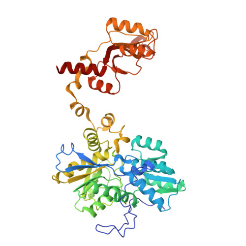
Human cystathionine beta-synthase (CBS) is a vital enzyme that regulates sulfur amino acid metabolism, hydrogen sulfide production, and cellular redox balance. Using a multidisciplinary approach, we demonstrate that CBS functions as a filamentous morpheein, with its stability, turnover, and activity governed by dynamic quaternary structural transitions. Three distinct filamentous assemblies were resolved by cryo-EM and are mediated by the oligomerization loop (residues 516-525): (i) ligand-free trans-dimers that form trans-basal filaments with basal stability and activity, (ii) adenosylornithine-bound cis-dimers that assemble into stabilized cis-basal filaments and (iii) S-adenosylmethionine-bound allo-dimers, which, together with cis-dimers, form highly stable, allo-activated stacked filaments. These reversible filamentous assemblies redefine CBS biology by integrating oligomerization and allosteric regulation within a morpheein framework. These findings provide a transformative perspective on CBS function and open avenues for pharmacological targeting of dysregulated CBS in various diseases including homocystinuria, cancer, and Down syndrome.
- Laboratory of Biological Electron Microscopy, Institute of Physics, School of Basic Sciences, Ecole Polytechnique Federale de Lausanne, Lausanne, Switzerland. inayathulla.mohammed@epfl.ch.
Organizational Affiliation: