9SPW | pdb_00009spw

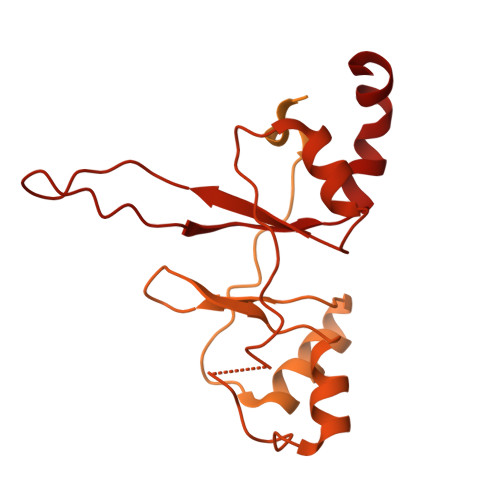
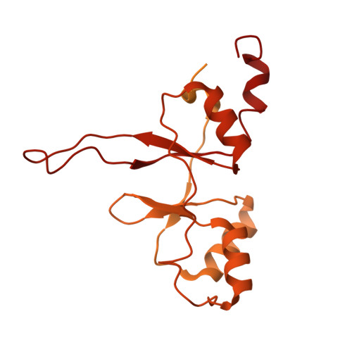
Structure of cis-basal conformer of human CBS induced by non-activating allosteric SAO ligand - by single particle approach.


Experimental Data Snapshot

  • Method: ELECTRON MICROSCOPY
  • Resolution: 4.11 Å
  • Aggregation State: PARTICLE 
  • Reconstruction Method: SINGLE PARTICLE 

wwPDB Validation 3D Report Full Report

Validation slider image for 9SPW

This is version 1.2 of the entry. See complete history. 

Literature

Structural basis for a filamentous morpheein model of human cystathionine beta-synthase.

Mohammed, I., Mijatovic, E., Philipp, T.M., Janickova, L., Ascencao, K., Asturias, F.J., Martinez-Cruz, L.A., Szabo, C., Stahlberg, H., Majtan, T.

(2026) Nat Commun 17

  • DOI: https://doi.org/10.1038/s41467-026-73198-7
  • Primary Citation Related Structures: 
    9SHM, 9SHN, 9SI8, 9SML, 9SPV, 9SPW, 9SQ0, 9SQQ

  • PubMed Abstract: 

    Human cystathionine beta-synthase (CBS) is a vital enzyme that regulates sulfur amino acid metabolism, hydrogen sulfide production, and cellular redox balance. Using a multidisciplinary approach, we demonstrate that CBS functions as a filamentous morpheein, with its stability, turnover, and activity governed by dynamic quaternary structural transitions. Three distinct filamentous assemblies were resolved by cryo-EM and are mediated by the oligomerization loop (residues 516-525): (i) ligand-free trans-dimers that form trans-basal filaments with basal stability and activity, (ii) adenosylornithine-bound cis-dimers that assemble into stabilized cis-basal filaments and (iii) S-adenosylmethionine-bound allo-dimers, which, together with cis-dimers, form highly stable, allo-activated stacked filaments. These reversible filamentous assemblies redefine CBS biology by integrating oligomerization and allosteric regulation within a morpheein framework. These findings provide a transformative perspective on CBS function and open avenues for pharmacological targeting of dysregulated CBS in various diseases including homocystinuria, cancer, and Down syndrome.


  • Organizational Affiliation: 
    • Laboratory of Biological Electron Microscopy, Institute of Physics, School of Basic Sciences, Ecole Polytechnique Federale de Lausanne, Lausanne, Switzerland. inayathulla.mohammed@epfl.ch.

Macromolecule Content 

  • Total Structure Weight: 368.65 kDa 
  • Atom Count: 12,194 
  • Modeled Residue Count: 1,554 
  • Deposited Residue Count: 3,306 
  • Unique protein chains: 2

Macromolecules

Find similar proteins by:|  3D Structure
Entity ID: 1
MoleculeChains  Sequence LengthOrganismDetailsImage
Cystathionine beta-synthase551Homo sapiensMutation(s): 0 
Gene Names: CBS
EC: 4.2.1.22
UniProt & NIH Common Fund Data Resources
Find proteins for P35520 (Homo sapiens)
Explore P35520 
Go to UniProtKB:  P35520
PHAROS:  P35520
GTEx:  ENSG00000160200 
Entity Groups
Sequence Clusters30% Identity50% Identity70% Identity90% Identity95% Identity100% Identity
UniProt GroupP35520
Sequence Annotations
Expand
Reference Sequence
Find similar proteins by:|  3D Structure
Entity ID: 2
MoleculeChains  Sequence LengthOrganismDetailsImage
Cystathionine beta-synthase
B, C, D, E, F
551Homo sapiensMutation(s): 0 
Gene Names: CBS
EC: 4.2.1.22
UniProt & NIH Common Fund Data Resources
Find proteins for P35520 (Homo sapiens)
Explore P35520 
Go to UniProtKB:  P35520
PHAROS:  P35520
GTEx:  ENSG00000160200 
Entity Groups
Sequence Clusters30% Identity50% Identity70% Identity90% Identity95% Identity100% Identity
UniProt GroupP35520
Sequence Annotations
Expand
Reference Sequence

Small Molecules

Ligands 2 Unique
IDChains Name / Formula / InChI Key2D Diagram3D Interactions
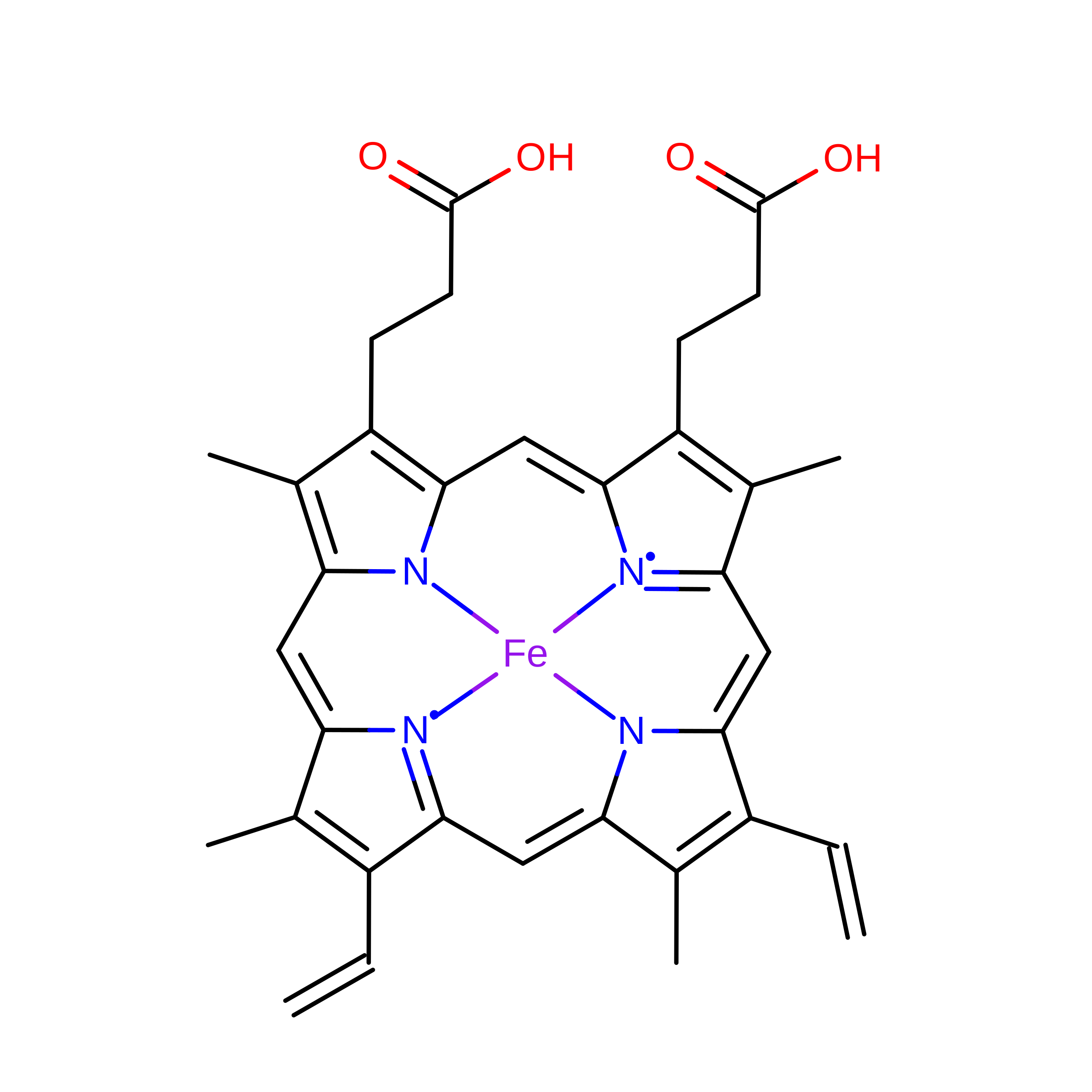
HEM
(Subject of Investigation/LOI)

Query on HEM


Download:Ideal Coordinates CCD File
I [auth C],
K [auth D]
PROTOPORPHYRIN IX CONTAINING FE
C34 H32 Fe N4 O4
KABFMIBPWCXCRK-RGGAHWMASA-L
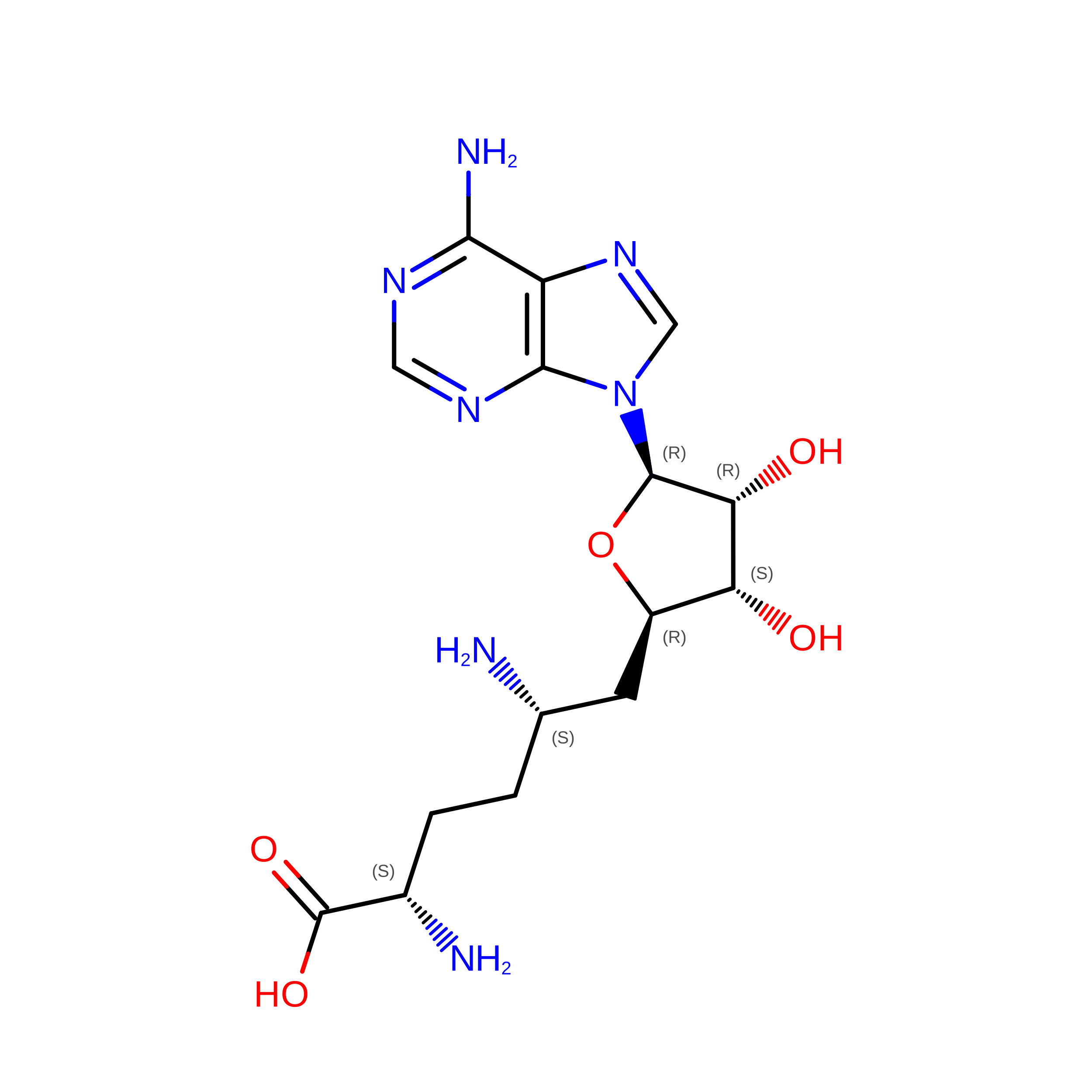
SFG
(Subject of Investigation/LOI)

Query on SFG


Download:Ideal Coordinates CCD File
G [auth A]
H [auth B]
J [auth C]
L [auth D]
M [auth E]
G [auth A],
H [auth B],
J [auth C],
L [auth D],
M [auth E],
N [auth F]
SINEFUNGIN
C15 H23 N7 O5
LMXOHSDXUQEUSF-YECHIGJVSA-N
Modified Residues  1 Unique
IDChains TypeFormula2D DiagramParent
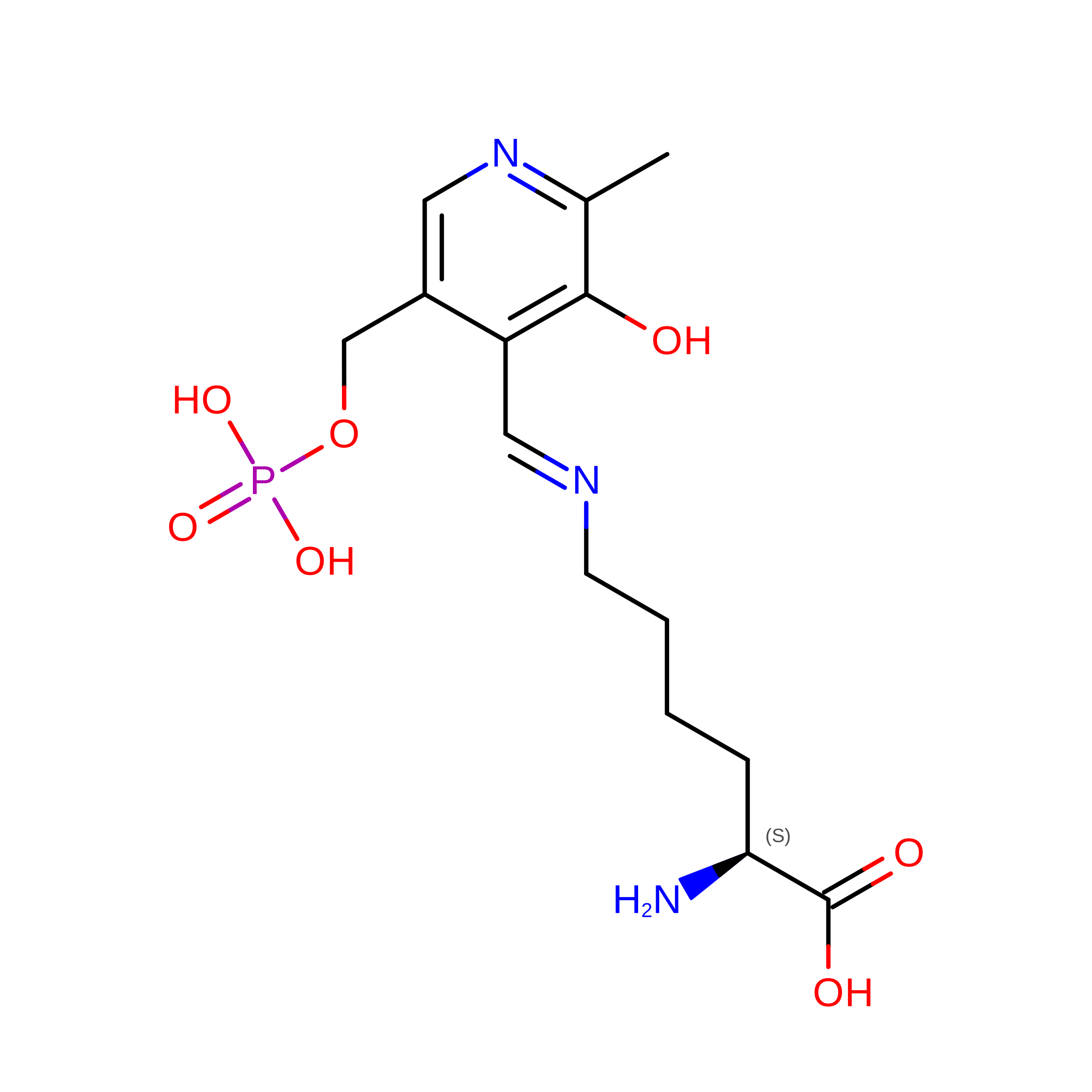
LLP
Query on LLP
B, C, D, E, F
L-PEPTIDE LINKINGC14 H22 N3 O7 PLYS

Experimental Data & Validation

Experimental Data

  • Method: ELECTRON MICROSCOPY
  • Resolution: 4.11 Å
  • Aggregation State: PARTICLE 
  • Reconstruction Method: SINGLE PARTICLE 
EM Software:
TaskSoftware PackageVersion
MODEL REFINEMENTPHENIXdev_5430
RECONSTRUCTIONcryoSPARC

Structure Validation

View Full Validation Report


Entry History 

& Funding Information

Deposition Data


Funding OrganizationLocationGrant Number
Swiss National Science FoundationSwitzerlandCRSII5_177195

Revision History  (Full details and data files)

  • Version 1.0: 2026-07-29
    Type: Initial release
  • Version 1.1: 2026-08-05
    Changes: Data collection, Database references
  • Version 1.2: 2026-09-02
    Type: Remediation
    Reason: Metalloprotein remediation
    Changes: Data collection, Derived calculations, Structure summary