9QHN | pdb_00009qhn

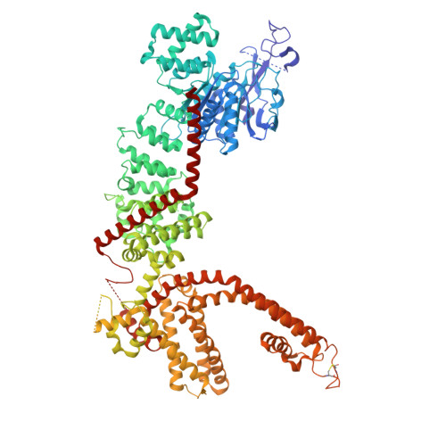
Cryo-EM structure of mouse TRPM3 alpha 2 in APO state


Experimental Data Snapshot

  • Method: ELECTRON MICROSCOPY
  • Resolution: 3.20 Å
  • Aggregation State: PARTICLE 
  • Reconstruction Method: SINGLE PARTICLE 

wwPDB Validation 3D Report Full Report

Validation slider image for 9QHN

This is version 1.1 of the entry. See complete history

Literature

Stereoselectivity and functional plasticity of a common ligand-binding pocket in TRPM3.

Bazeli, B.Shkumatov, A.V.Schenck, S.Vanherck, J.C.Janssens, A.Spieser, S.A.H.Marchand, D.Roelens, R.Chaltin, P.Marchand, A.Vriens, J.Voets, T.Brunner, J.D.

(2026) Nat Commun 17

  • DOI: https://doi.org/10.1038/s41467-026-71226-0
  • Primary Citation Related Structures: 
    9QHM, 9QHN, 9QHO, 9QHP, 9QHQ, 9T9U

  • PubMed Abstract: 

    The transient receptor potential melastatin 3 (TRPM3) channel is a key mediator of peripheral pain signaling, and pathogenic mutations in TRPM3 are linked to neurodevelopmental delay and epilepsy. Despite the therapeutic promise of TRPM3 modulators, the molecular mechanisms by which ligands modulate channel gating remain poorly understood. Here, we combine cryo-electron microscopy (cryo-EM) with functional analyses to characterize a promiscuous ligand-binding pocket formed by transmembrane helices S1-S4. This pocket accommodates several chemically diverse plant-derived and synthetic agonists and antagonists. We show stereoselectivity of TRPM3 for the (R)-enantiomer of the flavonoid antagonist isosakuranetin and the (R)-enantiomer of the synthetic agonist CIM0216. Mutations within this pocket-including variants identified in patients -alter ligand affinity and, in some cases, invert the functional outcome of ligand binding. These findings reveal the stereoselectivity and functional plasticity of the TRPM3 ligand-binding pocket, highlighting how subtle changes in the molecular interactions can produce divergent effects on channel gating, with important ramifications for TRPM3-targeted drug development and therapy.


  • Organizational Affiliation
    • Laboratory of Ion Channel Research (LICR), VIB-KU Leuven Center for Neuroscience, Leuven, Belgium, and Department of Molecular and Cellular Biology, KU Leuven, Leuven, Belgium.

Macromolecule Content 

  • Total Structure Weight: 548.95 kDa 
  • Atom Count: 32,684 
  • Modeled Residue Count: 3,984 
  • Deposited Residue Count: 4,708 
  • Unique protein chains: 1

Macromolecules

Find similar proteins by:|  3D Structure
Entity ID: 1
MoleculeChains  Sequence LengthOrganismDetailsImage
Transient receptor potential cation channel subfamily M member 3
A, B, C, D
1,177Mus musculusMutation(s): 0 
Gene Names: Trpm3
UniProt & NIH Common Fund Data Resources
Find proteins for J9SQF3 (Mus musculus)
Explore J9SQF3 
Go to UniProtKB:  J9SQF3
IMPC:  MGI:2443101
Entity Groups
Sequence Clusters30% Identity50% Identity70% Identity90% Identity95% Identity100% Identity
UniProt GroupJ9SQF3
Sequence Annotations
Expand
Reference Sequence

Small Molecules

Ligands 2 Unique
IDChains Name / Formula / InChI Key2D Diagram3D Interactions
3PH

Query on 3PH


Download:Ideal Coordinates CCD File
E [auth A],
G [auth B],
I [auth C],
K [auth D]
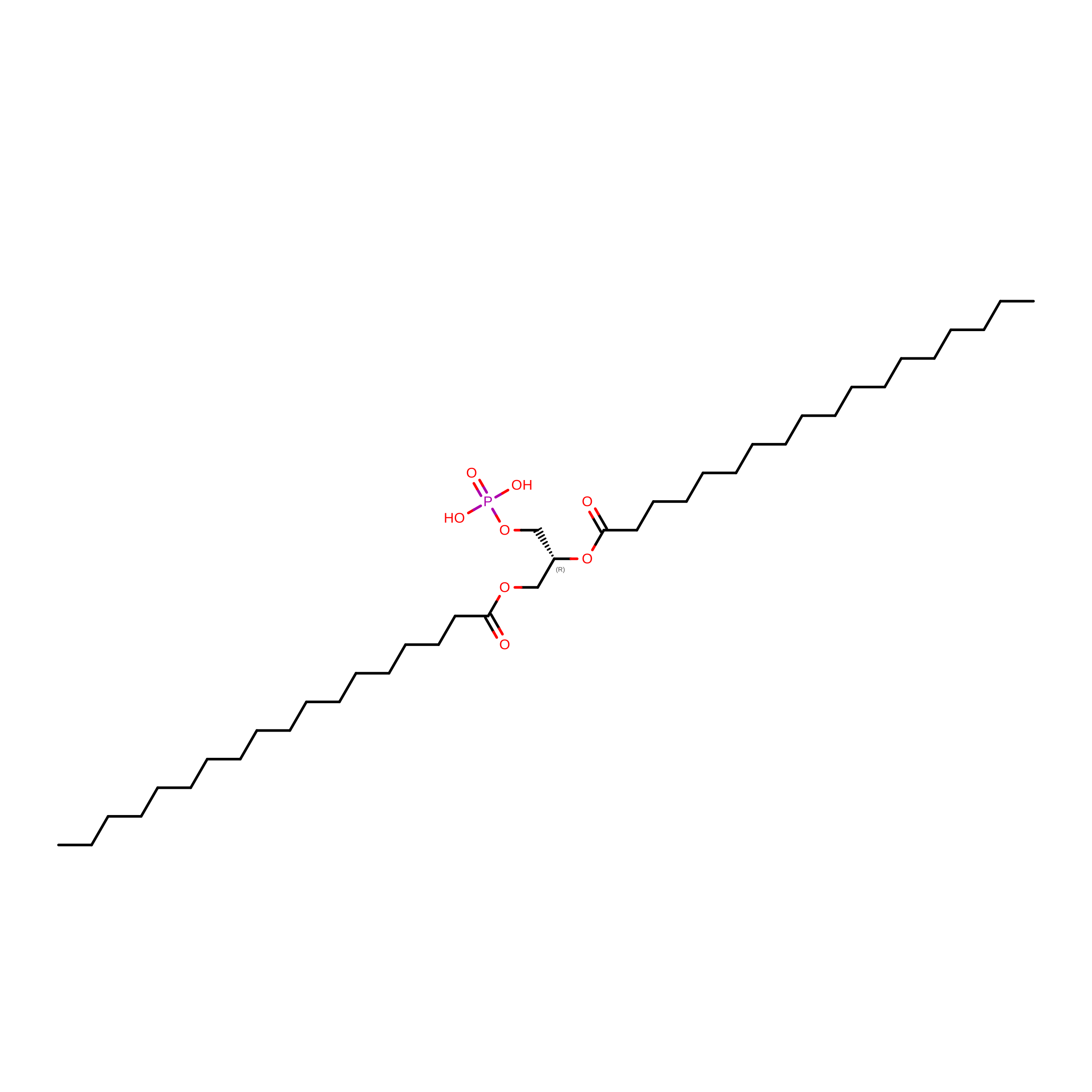
1,2-DIACYL-GLYCEROL-3-SN-PHOSPHATE
C39 H77 O8 P
YFWHNAWEOZTIPI-DIPNUNPCSA-N
CLR

Query on CLR


Download:Ideal Coordinates CCD File
F [auth A],
H [auth C],
J [auth D],
L [auth D]
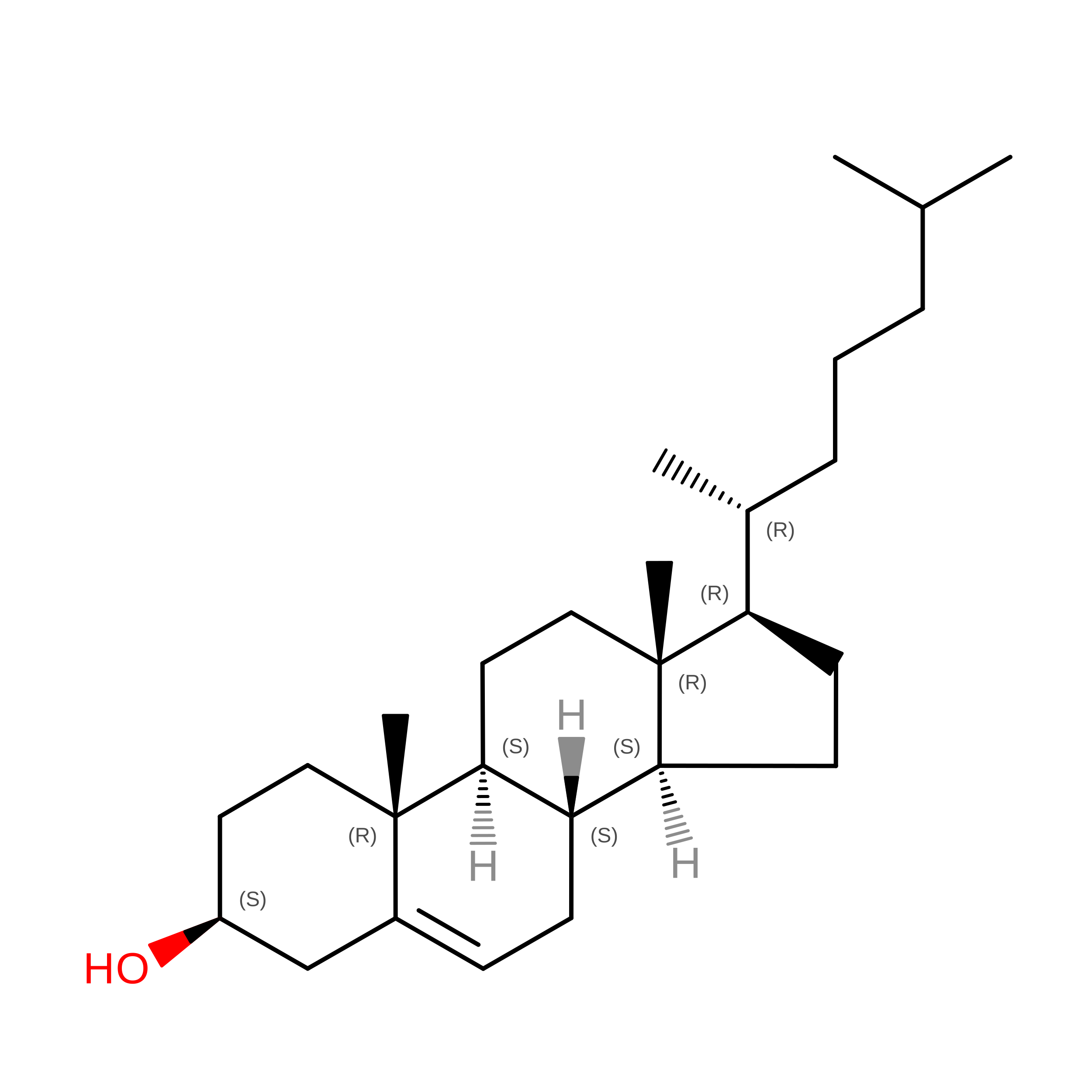
CHOLESTEROL
C27 H46 O
HVYWMOMLDIMFJA-DPAQBDIFSA-N

Experimental Data & Validation

Experimental Data

  • Method: ELECTRON MICROSCOPY
  • Resolution: 3.20 Å
  • Aggregation State: PARTICLE 
  • Reconstruction Method: SINGLE PARTICLE 
EM Software:
TaskSoftware PackageVersion
MODEL REFINEMENTPHENIX1.21-5207

Structure Validation

View Full Validation Report


Entry History 

& Funding Information

Deposition Data


Funding OrganizationLocationGrant Number
Not funded--

Revision History  (Full details and data files)

  • Version 1.0: 2026-02-11
    Type: Initial release
  • Version 1.1: 2026-08-26
    Changes: Data collection, Database references