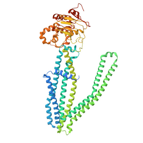
9NGJ | pdb_00009ngj

CryoEM structure of human ABCD3 bound to Phytanoyl-CoA


Experimental Data Snapshot

  • Method: ELECTRON MICROSCOPY
  • Resolution: 3.13 Å
  • Aggregation State: PARTICLE 
  • Reconstruction Method: SINGLE PARTICLE 

wwPDB Validation 3D Report Full Report

Validation slider image for 9NGJ

This is version 1.1 of the entry. See complete history

Literature

Molecular mechanism of substrate transport by human peroxisomal ABCD3.

Gupta, M.Khandelwal, N.K.Seka, D.J.Balasubramani, S.G.Dickinson, M.S.Myasnikov, A.Echeverria, I.Stroud, R.M.

(2025) Proc Natl Acad Sci U S A 122: e2513928122-e2513928122

  • DOI: https://doi.org/10.1073/pnas.2513928122
  • Primary Citation Related Structures: 
    9NGJ, 9NGM

  • PubMed Abstract: 

    ATP-binding cassette transporters of the D subfamily (ABCD1-3) mediate the export of CoA thioesters of fatty acids from the cytosol into peroxisomes for further oxidation. ABCD3 facilitates the transport of a broad spectrum of substrates, including branched-chain fatty acids, very long-chain fatty acids, bile salt intermediates, and dicarboxylic acids as CoA adducts. Mutations in ABCD3 are associated with defects in congenital bile acid synthesis. Despite its importance, the basis for substrate selectivity and the mechanism of transport by ABCD3 are not well defined. We report the cryogenic sample electron microscopy (cryo-EM) structures of full-length human ABCD3 in its apo state and bound to one of its physiological substrates (phytanoyl-CoA) at resolutions of 3.33 Å and 3.13 Å, respectively. Our biochemical assays reveal that substrate binding induces ATPase activity in ABCD3, suggesting a substrate-dependent conformational change. Structural comparison of the apo and substrate-bound states demonstrates that the substrate interaction brings nucleotide-binding domains closer together, providing a mechanistic basis of substrate-induced ATPase activity. These findings offer critical insights into the transport mechanism of ABCD3 and lay a structural foundation for understanding its role in peroxisomal metabolite import and related diseases.


  • Organizational Affiliation
    • Department of Chemical Physiology and Biochemistry, Oregon Health and Science University, Portland, OR 97239.

Macromolecule Content 

  • Total Structure Weight: 158.61 kDa 
  • Atom Count: 9,618 
  • Modeled Residue Count: 1,172 
  • Deposited Residue Count: 1,376 
  • Unique protein chains: 1

Macromolecules

Find similar proteins by:|  3D Structure
Entity ID: 1
MoleculeChains  Sequence LengthOrganismDetailsImage
ATP-binding cassette sub-family D member 3A [auth B],
B [auth A]
688Homo sapiensMutation(s): 0 
Gene Names: ABCD3PMP70PXMP1
EC: 3.1.2 (PDB Primary Data), 7.6.2 (PDB Primary Data)
UniProt & NIH Common Fund Data Resources
Find proteins for P28288 (Homo sapiens)
Explore P28288 
Go to UniProtKB:  P28288
GTEx:  ENSG00000117528 
Entity Groups
Sequence Clusters30% Identity50% Identity70% Identity90% Identity95% Identity100% Identity
UniProt GroupP28288
Sequence Annotations
Expand
Reference Sequence

Small Molecules

Ligands 1 Unique
IDChains Name / Formula / InChI Key2D Diagram3D Interactions
A1CAN
(Subject of Investigation/LOI)

Query on A1CAN


Download:Ideal Coordinates CCD File
C [auth B],
D [auth A]
S-{(3R,5S,9S)-1-[(2R,3S,4R,5R)-5-(6-amino-9H-purin-9-yl)-4-hydroxy-3-(phosphonooxy)oxolan-2-yl]-3,5,9-trihydroxy-8,8-dimethyl-3,5,10,14-tetraoxo-2,4,6-trioxa-11,15-diaza-3lambda~5~,5lambda~5~-diphosphaheptadecan-17-yl} (3S,7R,11R)-3,7,11,15-tetramethylhexadecanethioate (non-preferred name)
C41 H74 N7 O17 P3 S
NRJQGHHZMSOUEN-KBMDNNSFSA-N

Experimental Data & Validation

Experimental Data

  • Method: ELECTRON MICROSCOPY
  • Resolution: 3.13 Å
  • Aggregation State: PARTICLE 
  • Reconstruction Method: SINGLE PARTICLE 
EM Software:
TaskSoftware PackageVersion
MODEL REFINEMENTPHENIX1.21_5207
RECONSTRUCTIONcryoSPARC

Structure Validation

View Full Validation Report


Entry History 

& Funding Information

Deposition Data


Funding OrganizationLocationGrant Number
National Institutes of Health/National Institute on Aging (NIH/NIA)United States--

Revision History  (Full details and data files)

  • Version 1.0: 2025-12-17
    Type: Initial release
  • Version 1.1: 2026-07-29
    Changes: Data collection, Database references