Download Mendeley
Molecular mechanism of substrate transport by human peroxisomal ABCD3.
Gupta, M., Khandelwal, N.K., Seka, D.J., Balasubramani, S.G., Dickinson, M.S., Myasnikov, A., Echeverria, I., Stroud, R.M.(2025) Proc Natl Acad Sci U S A 122: e2513928122-e2513928122
- PubMed: 41428872 Search on PubMedSearch on PubMed Central
- DOI: https://doi.org/10.1073/pnas.2513928122
- Primary Citation Related Structures:
9NGJ, 9NGM - PubMed Abstract:
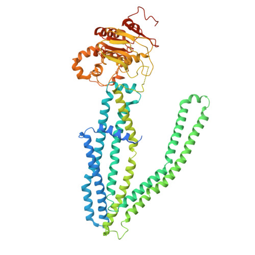
ATP-binding cassette transporters of the D subfamily (ABCD1-3) mediate the export of CoA thioesters of fatty acids from the cytosol into peroxisomes for further oxidation. ABCD3 facilitates the transport of a broad spectrum of substrates, including branched-chain fatty acids, very long-chain fatty acids, bile salt intermediates, and dicarboxylic acids as CoA adducts. Mutations in ABCD3 are associated with defects in congenital bile acid synthesis. Despite its importance, the basis for substrate selectivity and the mechanism of transport by ABCD3 are not well defined. We report the cryogenic sample electron microscopy (cryo-EM) structures of full-length human ABCD3 in its apo state and bound to one of its physiological substrates (phytanoyl-CoA) at resolutions of 3.33 Å and 3.13 Å, respectively. Our biochemical assays reveal that substrate binding induces ATPase activity in ABCD3, suggesting a substrate-dependent conformational change. Structural comparison of the apo and substrate-bound states demonstrates that the substrate interaction brings nucleotide-binding domains closer together, providing a mechanistic basis of substrate-induced ATPase activity. These findings offer critical insights into the transport mechanism of ABCD3 and lay a structural foundation for understanding its role in peroxisomal metabolite import and related diseases.
- Department of Chemical Physiology and Biochemistry, Oregon Health and Science University, Portland, OR 97239.
Organizational Affiliation: