 Download Mendeley
Dynamics of Class Ib ribonucleotide reductase complex revealed by CryoEM
Yadav, L.R., Mande, S.C., Vinothkumar, K.R., Kumar, J.To be published.
Experimental Data Snapshot
Starting Models: experimental
View more details
wwPDB Validation 3D Report Full Report

Entity ID: 1 | |||||
|---|---|---|---|---|---|
| Molecule | Chains | Sequence Length | Organism | Details | Image |
| Ribonucleoside-diphosphate reductase subunit beta | A, C [auth B] | 318 | Mycolicibacterium thermoresistibile ATCC 19527 | Mutation(s): 0 Gene Names: nrdF, KEK_07147 EC: 1.17.4.1 |  |
UniProt | |||||
Entity Groups | |||||
| Sequence Clusters | 30% Identity50% Identity70% Identity90% Identity95% Identity100% Identity | ||||
| UniProt Group | P9WH71 | ||||
Sequence AnnotationsExpand | |||||
Reference Sequence | |||||
Entity ID: 2 | |||||
|---|---|---|---|---|---|
| Molecule | Chains | Sequence Length | Organism | Details | Image |
| Ribonucleoside-diphosphate reductase | B [auth F], D [auth E] | 667 | Mycolicibacterium thermoresistibile ATCC 19527 | Mutation(s): 0 Gene Names: KEK_07092 EC: 1.17.4.1 |  |
UniProt | |||||
Entity Groups | |||||
| Sequence Clusters | 30% Identity50% Identity70% Identity90% Identity95% Identity100% Identity | ||||
| UniProt Group | P9WH75 | ||||
Sequence AnnotationsExpand | |||||
Reference Sequence | |||||
| Ligands 1 Unique | |||||
|---|---|---|---|---|---|
| ID | Chains | Name / Formula / InChI Key | 2D Diagram | 3D Interactions | |
| MN (Subject of Investigation/LOI) Download:Ideal Coordinates CCD File | E [auth A], F [auth A], G [auth B], H [auth B] | MANGANESE (II) ION Mn WAEMQWOKJMHJLA-UHFFFAOYSA-N | 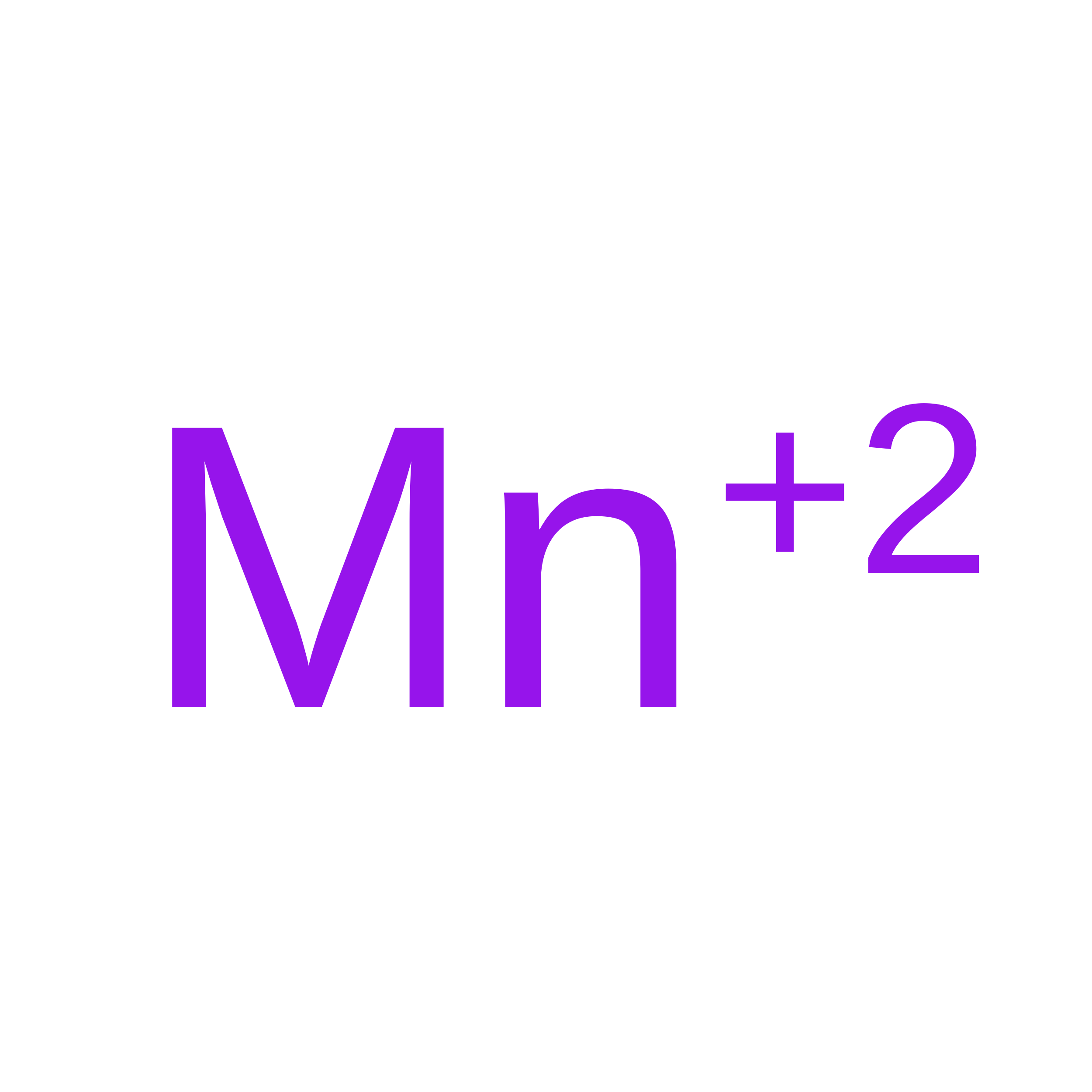 | ||
| Task | Software Package | Version |
|---|---|---|
| RECONSTRUCTION | cryoSPARC | v3.3.2 |
| MODEL REFINEMENT | PHENIX | 1.20.1_4487 |
| Funding Organization | Location | Grant Number |
|---|---|---|
| Department of Biotechnology (DBT, India) | India | Centre of Excellence Grant (BT/PR15450/COE/34/46/2016) |
| Department of Biotechnology (DBT, India) | India | DBT/PR12422/MED/31/287/2014 |
