9JOW | pdb_00009jow

Cryo-EM structure of the myxol-bound light-driven chloride ion-pumping rhodopsin, NM-R3


Experimental Data Snapshot

  • Method: ELECTRON MICROSCOPY
  • Resolution: 2.48 Å
  • Aggregation State: PARTICLE 
  • Reconstruction Method: SINGLE PARTICLE 

wwPDB Validation   3D Report Full Report


This is version 1.1 of the entry. See complete history


Literature

Carotenoids bind rhodopsins and act as photocycle-accelerating pigments in marine Bacteroidota.

Fujiwara, T.Hosaka, T.Hasegawa-Takano, M.Nishimura, Y.Tominaga, K.Mori, K.Nishino, S.Takahashi, Y.Uchikubo-Kamo, T.Hanada, K.Maoka, T.Takaichi, S.Inoue, K.Shirouzu, M.Yoshizawa, S.

(2025) Nat Microbiol 10: 2603-2615

  • DOI: https://doi.org/10.1038/s41564-025-02109-1
  • Primary Citation of Related Structures:  
    9JOU, 9JOV, 9JOW, 9JOX

  • PubMed Abstract: 

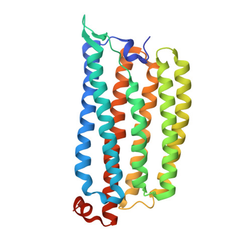
    Microbial rhodopsins are photoreceptor proteins widely distributed in marine microorganisms that harness light energy and support marine ecosystems. While retinal is typically the sole chromophore in microbial rhodopsins, some proteorhodopsins, which are proton-pumping rhodopsins abundant in the ocean, use carotenoid antennae to transfer light energy to retinal. However, the mechanism by which carotenoids enhance rhodopsin functions remains unclear. Here, using the marine Bacteroidota isolate Nonlabens marinus S1-08 T , we reconstituted complexes of rhodopsins with the carotenoid myxol and detected energy transfer to retinal in both proteorhodopsin and chloride ion-pumping rhodopsin. Carotenoid binding facilitated light harvesting and accelerated the photocycle, thereby improving the light utilization efficiency of proteorhodopsin. Cryogenic electron microscopy structural analysis further revealed the molecular architecture of the carotenoid-rhodopsin complexes. The ability to bind carotenoids is conserved in rhodopsins of the marine-dominant phylum Bacteroidota, which are widely transcribed in the photic zone. These findings reveal how carotenoids enhance rhodopsin functions in marine Bacteroidota.


  • Organizational Affiliation
    • Graduate School of Frontier Sciences, The University of Tokyo, Chiba, Japan.

Macromolecules
Find similar proteins by:  (by identity cutoff)  |  3D Structure
Entity ID: 1
MoleculeChains Sequence LengthOrganismDetailsImage
Chloride pumping rhodopsin
A, B, C, D, E
279Nonlabens marinus S1-08Mutation(s): 0 
Gene Names: ClRNMS_1267
UniProt
Find proteins for W8VZW3 (Nonlabens marinus S1-08)
Explore W8VZW3 
Go to UniProtKB:  W8VZW3
Entity Groups  
Sequence Clusters30% Identity50% Identity70% Identity90% Identity95% Identity100% Identity
UniProt GroupW8VZW3
Sequence Annotations
Expand
  • Reference Sequence
Small Molecules
Ligands 10 Unique
IDChains Name / Formula / InChI Key2D Diagram3D Interactions
PC1 (Subject of Investigation/LOI)
Query on PC1

Download Ideal Coordinates CCD File 
CB [auth E],
HA [auth C],
J [auth A],
SA [auth D],
W [auth B]
1,2-DIACYL-SN-GLYCERO-3-PHOSPHOCHOLINE
C44 H88 N O8 P
NRJAVPSFFCBXDT-HUESYALOSA-N
PLC (Subject of Investigation/LOI)
Query on PLC

Download Ideal Coordinates CCD File 
IA [auth C],
K [auth A],
P [auth A],
TA [auth D],
X [auth B]
DIUNDECYL PHOSPHATIDYL CHOLINE
C32 H65 N O8 P
IJFVSSZAOYLHEE-SSEXGKCCSA-O
A1L4O
Query on A1L4O

Download Ideal Coordinates CCD File 
EA [auth C],
G [auth A],
PA [auth D],
T [auth B],
ZA [auth E]
(3~{S},4~{Z},6~{E},8~{Z},10~{E},12~{E},14~{E},16~{E},18~{E},20~{E},22~{E},24~{E})-2,6,10,14,19,23-hexamethyl-25-[(4~{R})-2,6,6-trimethyl-4-oxidanyl-cyclohexen-1-yl]pentacosa-4,6,8,10,12,14,16,18,20,22,24-undecaene-2,3-diol
C40 H56 O3
JJPMVSRTRMLHST-CNFSYYPWSA-N
RET (Subject of Investigation/LOI)
Query on RET

Download Ideal Coordinates CCD File 
DA [auth C],
F [auth A],
OA [auth D],
S [auth B],
YA [auth E]
RETINAL
C20 H28 O
NCYCYZXNIZJOKI-OVSJKPMPSA-N
8K6 (Subject of Investigation/LOI)
Query on 8K6

Download Ideal Coordinates CCD File 
MA [auth C],
N [auth A],
R [auth B]
Octadecane
C18 H38
RZJRJXONCZWCBN-UHFFFAOYSA-N
R16 (Subject of Investigation/LOI)
Query on R16

Download Ideal Coordinates CCD File 
AA [auth B]
BA [auth B]
FB [auth E]
L [auth A]
M [auth A]
AA [auth B],
BA [auth B],
FB [auth E],
L [auth A],
M [auth A],
NA [auth D],
Q [auth A],
UA [auth D],
VA [auth D],
XA [auth E],
Z [auth B]
HEXADECANE
C16 H34
DCAYPVUWAIABOU-UHFFFAOYSA-N
C14 (Subject of Investigation/LOI)
Query on C14

Download Ideal Coordinates CCD File 
CA [auth B]
EB [auth E]
GB [auth E]
JA [auth C]
KA [auth C]
CA [auth B],
EB [auth E],
GB [auth E],
JA [auth C],
KA [auth C],
Y [auth B]
TETRADECANE
C14 H30
BGHCVCJVXZWKCC-UHFFFAOYSA-N
D12 (Subject of Investigation/LOI)
Query on D12

Download Ideal Coordinates CCD File 
DB [auth E],
O [auth A],
WA [auth D]
DODECANE
C12 H26
SNRUBQQJIBEYMU-UHFFFAOYSA-N
D10 (Subject of Investigation/LOI)
Query on D10

Download Ideal Coordinates CCD File 
LA [auth C]DECANE
C10 H22
DIOQZVSQGTUSAI-UHFFFAOYSA-N
CL (Subject of Investigation/LOI)
Query on CL

Download Ideal Coordinates CCD File 
AB [auth E]
BB [auth E]
FA [auth C]
GA [auth C]
H [auth A]
AB [auth E],
BB [auth E],
FA [auth C],
GA [auth C],
H [auth A],
I [auth A],
QA [auth D],
RA [auth D],
U [auth B],
V [auth B]
CHLORIDE ION
Cl
VEXZGXHMUGYJMC-UHFFFAOYSA-M
Experimental Data & Validation

Experimental Data

  • Method: ELECTRON MICROSCOPY
  • Resolution: 2.48 Å
  • Aggregation State: PARTICLE 
  • Reconstruction Method: SINGLE PARTICLE 

Structure Validation

View Full Validation Report


Entry History & Funding Information

Deposition Data


Funding OrganizationLocationGrant Number
Japan Society for the Promotion of Science (JSPS)Japan--

Revision History  (Full details and data files)

  • Version 1.0: 2025-07-30
    Type: Initial release
  • Version 1.1: 2026-02-18
    Changes: Data collection, Database references