9IZG | pdb_00009izg

Cryo-EM structure of LPA1-Gq complex with LPA


Experimental Data Snapshot

  • Method: ELECTRON MICROSCOPY
  • Resolution: 3.04 Å
  • Aggregation State: PARTICLE 
  • Reconstruction Method: SINGLE PARTICLE 

wwPDB Validation 3D Report Full Report

Validation slider image for 9IZG

This is version 1.1 of the entry. See complete history

Literature

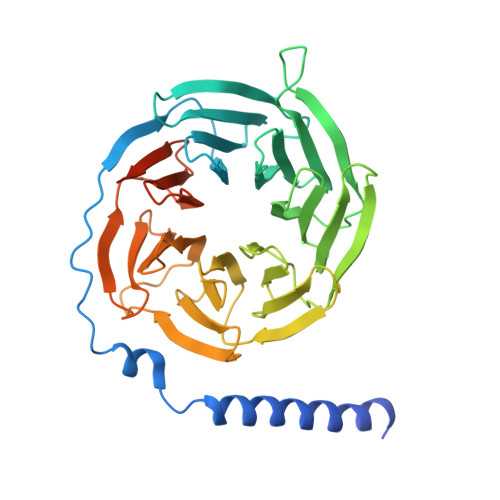
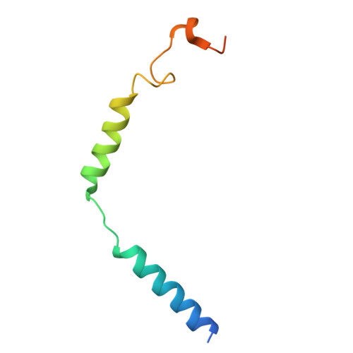
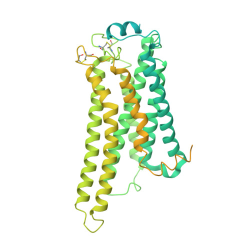
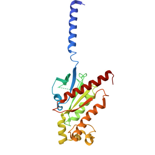
Structural insights into the engagement of lysophosphatidic acid receptor 1 with different G proteins.

Suzuki, S.Tanaka, K.Kamegawa, A.Nishikawa, K.Suzuki, H.Oshima, A.Fujiyoshi, Y.

(2024) J Struct Biol 217: 108164-108164

  • DOI: https://doi.org/10.1016/j.jsb.2024.108164
  • Primary Citation Related Structures: 
    9IZF, 9IZG, 9IZH

  • PubMed Abstract: 

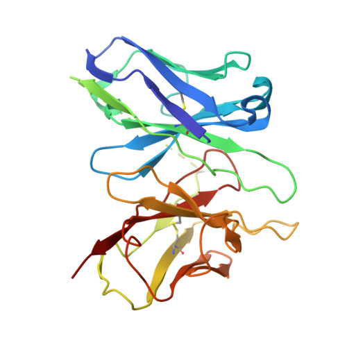
    Lysophosphatidic acid (LPA) and sphingosine-1-phosphate (S1P) are bioactive lysophospholipids derived from cell membranes that activate the endothelial differentiation gene family of G protein-coupled receptors. Activation of these receptors triggers multiple downstream signaling cascades through G proteins such as Gi/o, Gq/11, and G12/13. Therefore, LPA and S1P mediate several physiological processes, including cytoskeletal dynamics, neurite retraction, cell migration, cell proliferation, and intracellular ion fluxes. The basis for the G-protein coupling selectivity of EDG receptors, however, remains unknown. Here, we present cryo-electron microscopy structures of LPA-activated LPA1 in complexes with G i , G q , and G 13 heterotrimers . Comparison of the three LPA1-G protein structures shows clearly different conformations of intracellular loop 2 (ICL2) and ICL3 that are likely induced by the different Gα protein interfaces. Interestingly, this G-protein interface interaction is a common feature of LPA and S1P receptors. Our findings provide clues to understanding the promiscuity of G-protein coupling in the endothelial differentiation gene family.


  • Organizational Affiliation
    • Advanced Research Initiative, Institute of Integrated Research, Institute of Science Tokyo, 1-5-45 Yushima Bunkyo-ku 113-8510, Tokyo, Japan.

Macromolecule Content 

  • Total Structure Weight: 177.68 kDa 
  • Atom Count: 8,702 
  • Modeled Residue Count: 1,149 
  • Deposited Residue Count: 1,597 
  • Unique protein chains: 5

Macromolecules

Find similar proteins by:|  3D Structure
Entity ID: 1
MoleculeChains  Sequence LengthOrganismDetailsImage
Guanine nucleotide-binding protein G(I)/G(S)/G(T) subunit beta-1A [auth B]382Homo sapiensMutation(s): 0 
Gene Names: GNB1
UniProt & NIH Common Fund Data Resources
Find proteins for P62873 (Homo sapiens)
Explore P62873 
Go to UniProtKB:  P62873
PHAROS:  P62873
GTEx:  ENSG00000078369 
Entity Groups
Sequence Clusters30% Identity50% Identity70% Identity90% Identity95% Identity100% Identity
UniProt GroupP62873
Sequence Annotations
Expand
Reference Sequence
Find similar proteins by:|  3D Structure
Entity ID: 2
MoleculeChains  Sequence LengthOrganismDetailsImage
Guanine nucleotide-binding protein G(I)/G(S)/G(O) subunit gamma-2B [auth C]70Mus musculusMutation(s): 0 
Gene Names: Gng2
UniProt
Find proteins for P63213 (Mus musculus)
Explore P63213 
Go to UniProtKB:  P63213
Entity Groups
Sequence Clusters30% Identity50% Identity70% Identity90% Identity95% Identity100% Identity
UniProt GroupP63213
Sequence Annotations
Expand
Reference Sequence
Find similar proteins by:|  3D Structure
Entity ID: 3
MoleculeChains  Sequence LengthOrganismDetailsImage
Soluble cytochrome b562,Lysophosphatidic acid receptor 1,LgBiT tagC [auth R]651Escherichia coliHomo sapienssynthetic construct
This entity is chimeric
Mutation(s): 0 
Gene Names: cybCLPAR1EDG2LPA1
UniProt & NIH Common Fund Data Resources
Find proteins for P0ABE7 (Escherichia coli)
Explore P0ABE7 
Go to UniProtKB:  P0ABE7
Find proteins for Q92633 (Homo sapiens)
Explore Q92633 
Go to UniProtKB:  Q92633
PHAROS:  Q92633
GTEx:  ENSG00000198121 
Entity Groups
Sequence Clusters30% Identity50% Identity70% Identity90% Identity95% Identity100% Identity
UniProt GroupsP0ABE7Q92633
Sequence Annotations
Expand
Reference Sequence
Find similar proteins by:|  3D Structure
Entity ID: 4
MoleculeChains  Sequence LengthOrganismDetailsImage
Gs-mini-Gq chimeraD [auth A]246Homo sapiensMutation(s): 0 
UniProt & NIH Common Fund Data Resources
Find proteins for A0A590UJY2 (Homo sapiens)
Explore A0A590UJY2 
Go to UniProtKB:  A0A590UJY2
GTEx:  ENSG00000087460 
Entity Groups
Sequence Clusters30% Identity50% Identity70% Identity90% Identity95% Identity100% Identity
UniProt GroupA0A590UJY2
Sequence Annotations
Expand
Reference Sequence
Find similar proteins by:|  3D Structure
Entity ID: 5
MoleculeChains  Sequence LengthOrganismDetailsImage
scFv16E [auth S]248synthetic constructMutation(s): 0 

Small Molecules

Ligands 1 Unique
IDChains Name / Formula / InChI Key2D Diagram3D Interactions
NKP
(Subject of Investigation/LOI)

Query on NKP


Download:Ideal Coordinates CCD File
F [auth R](2R)-2-hydroxy-3-(phosphonooxy)propyl (9E)-octadec-9-enoate
C21 H41 O7 P
WRGQSWVCFNIUNZ-SQUSKLHYSA-N

Experimental Data & Validation

Experimental Data

  • Method: ELECTRON MICROSCOPY
  • Resolution: 3.04 Å
  • Aggregation State: PARTICLE 
  • Reconstruction Method: SINGLE PARTICLE 

Structure Validation

View Full Validation Report


Entry History 

& Funding Information

Deposition Data


Funding OrganizationLocationGrant Number
Japan Society for the Promotion of Science (JSPS)Japan20H00451

Revision History  (Full details and data files)

  • Version 1.0: 2025-01-01
    Type: Initial release
  • Version 1.1: 2025-01-15
    Changes: Data collection, Database references