9HX3 | pdb_00009hx3

Amyloid fibril of TTR


Experimental Data Snapshot

  • Method: ELECTRON MICROSCOPY
  • Resolution: 3.00 Å
  • Aggregation State: FILAMENT 
  • Reconstruction Method: HELICAL 

wwPDB Validation 3D Report Full Report

Validation slider image for 9HX3

This is version 1.0 of the entry. See complete history

Literature

Apolipoprotein A-IV fibrils: structural diagnosis of mixed cardiac amyloidosis.

Aibara, S.Kassner, A.Wong, E.Klingel, K.Papworth, M.Althage, M.Wang, Q.D.Correia, C.Milting, H.de Oliveira, T.M.

(2025) Nat Commun 16: 9276-9276

  • DOI: https://doi.org/10.1038/s41467-025-64902-0
  • Primary Citation Related Structures: 
    9HX3, 9HX4

  • PubMed Abstract: 

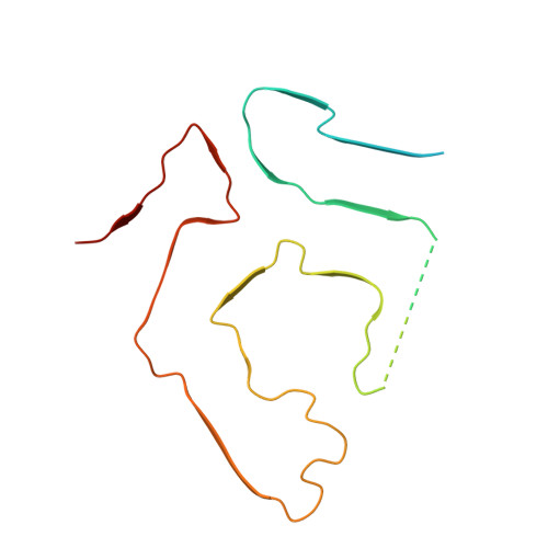
    Cardiac amyloidosis (CA) occurs when misfolded proteins deposit as fibrils in the extracellular space of the heart. The fibrillogenic properties of apolipoprotein A-IV (ApoAIV) have been histologically observed and associated with CA pathogenesis. We report the structure of an ApoAIV amyloid from a patient's heart, which coexist amongst transthyretin (TTR) amyloids. These cases of undetected mixed CA highlight the importance of developing broad-spectrum anti-amyloid treatments to improve outcomes in patients.


  • Organizational Affiliation
    • Protein Sciences, Structure, and Biophysics, Discovery Sciences, R&D, AstraZeneca, Cambridge, UK.

Macromolecule Content 

  • Total Structure Weight: 79.52 kDa 
  • Atom Count: 3,630 
  • Modeled Residue Count: 465 
  • Deposited Residue Count: 735 
  • Unique protein chains: 1

Macromolecules

Find similar proteins by:|  3D Structure
Entity ID: 1
MoleculeChains  Sequence LengthOrganismDetailsImage
TransthyretinA,
B [auth C],
C [auth B],
D [auth E],
E [auth D]
147Homo sapiensMutation(s): 0 
UniProt & NIH Common Fund Data Resources
Find proteins for P02766 (Homo sapiens)
Explore P02766 
Go to UniProtKB:  P02766
PHAROS:  P02766
GTEx:  ENSG00000118271 
Entity Groups
Sequence Clusters30% Identity50% Identity70% Identity90% Identity95% Identity100% Identity
UniProt GroupP02766
Sequence Annotations
Expand
Reference Sequence

Experimental Data & Validation

Experimental Data

  • Method: ELECTRON MICROSCOPY
  • Resolution: 3.00 Å
  • Aggregation State: FILAMENT 
  • Reconstruction Method: HELICAL 
EM Software:
TaskSoftware PackageVersion
MODEL REFINEMENTPHENIX1.21.1_5286:

Structure Validation

View Full Validation Report


Entry History 

& Funding Information

Deposition Data


Funding OrganizationLocationGrant Number
Not funded--

Revision History  (Full details and data files)

  • Version 1.0: 2025-11-05
    Type: Initial release