9FJU

Structure of the DNase I- and phalloidin-bound pointed end of F-actin (conformer 1)


Experimental Data Snapshot

  • Method: ELECTRON MICROSCOPY
  • Resolution: 3.84 Å
  • Aggregation State: PARTICLE 
  • Reconstruction Method: SINGLE PARTICLE 

Starting Models: experimental
View more details

wwPDB Validation   3D Report Full Report


This is version 1.1 of the entry. See complete history


Literature

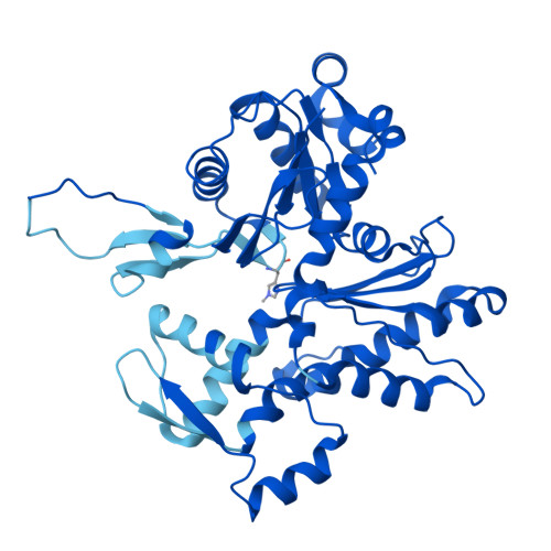
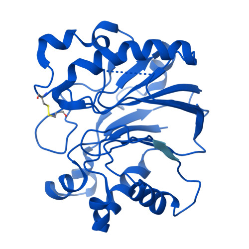
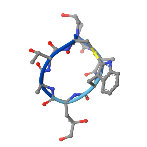
Phalloidin and DNase I-bound F-actin pointed end structures reveal principles of filament stabilization and disassembly.

Boiero Sanders, M.Oosterheert, W.Hofnagel, O.Bieling, P.Raunser, S.

(2024) Nat Commun 15: 7969-7969

  • DOI: https://doi.org/10.1038/s41467-024-52251-3
  • Primary Citation of Related Structures:  
    9FJM, 9FJO, 9FJU, 9FJY

  • PubMed Abstract: 

    Actin filament turnover involves subunits binding to and dissociating from the filament ends, with the pointed end being the primary site of filament disassembly. Several molecules modulate filament turnover, but the underlying mechanisms remain incompletely understood. Here, we present three cryo-EM structures of the F-actin pointed end in the presence and absence of phalloidin or DNase I. The two terminal subunits at the undecorated pointed end adopt a twisted conformation. Phalloidin can still bind and bridge these subunits, inducing a conformational shift to a flattened, F-actin-like state. This explains how phalloidin prevents depolymerization at the pointed end. Interestingly, two DNase I molecules simultaneously bind to the phalloidin-stabilized pointed end. In the absence of phalloidin, DNase I binding would disrupt the terminal actin subunit packing, resulting in filament disassembly. Our findings uncover molecular principles of pointed end regulation and provide structural insights into the kinetic asymmetry between the actin filament ends.


  • Organizational Affiliation

    Department of Structural Biochemistry, Max Planck Institute of Molecular Physiology, 44227, Dortmund, Germany.


Macromolecules
Find similar proteins by:  (by identity cutoff)  |  3D Structure
Entity ID: 1
MoleculeChains Sequence LengthOrganismDetailsImage
Actin, cytoplasmic 1, N-terminally processed
A, B, C, D
374Bos taurusMutation(s): 0 
UniProt
Find proteins for P60712 (Bos taurus)
Explore P60712 
Go to UniProtKB:  P60712
Entity Groups  
Sequence Clusters30% Identity50% Identity70% Identity90% Identity95% Identity100% Identity
UniProt GroupP60712
Sequence Annotations
Expand
  • Reference Sequence
Find similar proteins by:  (by identity cutoff)  |  3D Structure
Entity ID: 2
MoleculeChains Sequence LengthOrganismDetailsImage
Deoxyribonuclease-1
E, F
282Bos taurusMutation(s): 0 
EC: 3.1.21.1
UniProt
Find proteins for P00639 (Bos taurus)
Explore P00639 
Go to UniProtKB:  P00639
Entity Groups  
Sequence Clusters30% Identity50% Identity70% Identity90% Identity95% Identity100% Identity
UniProt GroupP00639
Glycosylation
Glycosylation Sites: 1
Sequence Annotations
Expand
  • Reference Sequence

Find similar proteins by:  Sequence   |   3D Structure  

Entity ID: 3
MoleculeChains Sequence LengthOrganismDetailsImage
Phalloidin
G, H, I, J
7Amanita phalloidesMutation(s): 0 
Sequence Annotations
Expand
  • Reference Sequence
Oligosaccharides

Help

Entity ID: 4
MoleculeChains Length2D Diagram Glycosylation3D Interactions
2-acetamido-2-deoxy-beta-D-glucopyranose-(1-4)-2-acetamido-2-deoxy-beta-D-glucopyranose
K, L
2N-Glycosylation
Glycosylation Resources
GlyTouCan:  G42666HT
GlyCosmos:  G42666HT
GlyGen:  G42666HT
Small Molecules
Ligands 3 Unique
IDChains Name / Formula / InChI Key2D Diagram3D Interactions
ADP (Subject of Investigation/LOI)
Query on ADP

Download Ideal Coordinates CCD File 
M [auth A],
P [auth B],
S [auth C],
V [auth D]
ADENOSINE-5'-DIPHOSPHATE
C10 H15 N5 O10 P2
XTWYTFMLZFPYCI-KQYNXXCUSA-N
PO4 (Subject of Investigation/LOI)
Query on PO4

Download Ideal Coordinates CCD File 
O [auth A],
R [auth B],
U [auth C],
X [auth D]
PHOSPHATE ION
O4 P
NBIIXXVUZAFLBC-UHFFFAOYSA-K
MG
Query on MG

Download Ideal Coordinates CCD File 
AA [auth F]
BA [auth F]
N [auth A]
Q [auth B]
T [auth C]
AA [auth F],
BA [auth F],
N [auth A],
Q [auth B],
T [auth C],
W [auth D],
Y [auth E],
Z [auth E]
MAGNESIUM ION
Mg
JLVVSXFLKOJNIY-UHFFFAOYSA-N
Modified Residues  2 Unique
IDChains TypeFormula2D DiagramParent
HIC
Query on HIC
A, B, C, D
L-PEPTIDE LINKINGC7 H11 N3 O2HIS
HYP
Query on HYP
G, H, I, J
L-PEPTIDE LINKINGC5 H9 N O3PRO
Biologically Interesting Molecules (External Reference) 1 Unique
Experimental Data & Validation

Experimental Data

  • Method: ELECTRON MICROSCOPY
  • Resolution: 3.84 Å
  • Aggregation State: PARTICLE 
  • Reconstruction Method: SINGLE PARTICLE 
EM Software:
TaskSoftware PackageVersion
RECONSTRUCTIONcryoSPARCv4.2.1
MODEL REFINEMENTCoot0.9.8.1
MODEL REFINEMENTPHENIX1.21rc1_5015

Structure Validation

View Full Validation Report


Entry History & Funding Information

Deposition Data


Funding OrganizationLocationGrant Number
Alexander von Humboldt FoundationGermany--
German Research Foundation (DFG)GermanyBI 1998/2-1
European Research Council (ERC)European Union856118

Revision History  (Full details and data files)

  • Version 1.0: 2024-09-11
    Type: Initial release
  • Version 1.1: 2024-10-02
    Changes: Data collection, Database references