9FAZ

Gcase in complex with small molecule inhibitor 1


Experimental Data Snapshot

  • Method: X-RAY DIFFRACTION
  • Resolution: 1.63 Å
  • R-Value Free: 0.195 
  • R-Value Work: 0.159 
  • R-Value Observed: 0.160 

wwPDB Validation   3D Report Full Report


Ligand Structure Quality Assessment 


This is version 1.1 of the entry. See complete history


Literature

Fragment-Based Discovery of a Series of Allosteric-Binding Site Modulators of beta-Glucocerebrosidase.

Palmer, N.Agnew, C.Benn, C.Buffham, W.J.Castro, J.N.Chessari, G.Clark, M.Cons, B.D.Coyle, J.E.Dawson, L.A.Hamlett, C.C.F.Hodson, C.Holding, F.Johnson, C.N.Liebeschuetz, J.W.Mahajan, P.McCarthy, J.M.Murray, C.W.O'Reilly, M.Peakman, T.Price, A.Rapti, M.Reeks, J.Schopf, P.St-Denis, J.D.Valenzano, C.Wallis, N.G.Walser, R.Weir, H.Wilsher, N.E.Woodhead, A.Bento, C.F.Tisi, D.

(2024) J Med Chem 67: 11168-11181

  • DOI: https://doi.org/10.1021/acs.jmedchem.4c00702
  • Primary Citation of Related Structures:  
    9F9Z, 9FA3, 9FA6, 9FAD, 9FAL, 9FAY, 9FAZ, 9FB2, 9FDI

  • PubMed Abstract: 

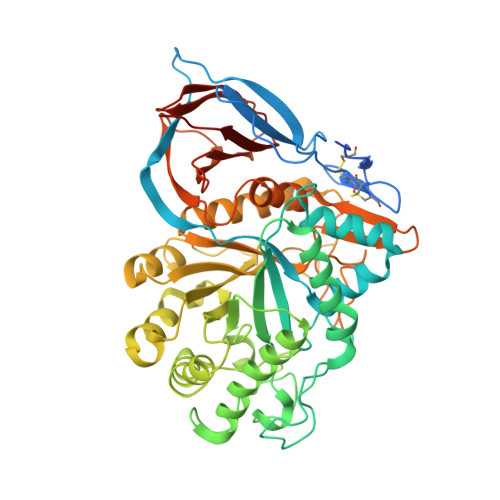
    β-Glucocerebrosidase (GBA/GCase) mutations leading to misfolded protein cause Gaucher's disease and are a major genetic risk factor for Parkinson's disease and dementia with Lewy bodies. The identification of small molecule pharmacological chaperones that can stabilize the misfolded protein and increase delivery of degradation-prone mutant GCase to the lysosome is a strategy under active investigation. Here, we describe the first use of fragment-based drug discovery (FBDD) to identify pharmacological chaperones of GCase. The fragment hits were identified by using X-ray crystallography and biophysical techniques. This work led to the discovery of a series of compounds that bind GCase with nM potency and positively modulate GCase activity in cells.


  • Organizational Affiliation

    Astex Pharmaceuticals, 436 Cambridge Science Park, Milton Road, Cambridge CB4 0QA, U.K.


Macromolecules
Find similar proteins by:  (by identity cutoff)  |  3D Structure
Entity ID: 1
MoleculeChains Sequence LengthOrganismDetailsImage
Lysosomal acid glucosylceramidase546Homo sapiensMutation(s): 0 
Gene Names: GBA1GBAGCGLUC
EC: 3.2.1.45 (PDB Primary Data), 2.4.1 (PDB Primary Data), 3.2.1 (PDB Primary Data), 3.2.1.46 (PDB Primary Data)
UniProt & NIH Common Fund Data Resources
Find proteins for P04062 (Homo sapiens)
Explore P04062 
Go to UniProtKB:  P04062
GTEx:  ENSG00000177628 
Entity Groups  
Sequence Clusters30% Identity50% Identity70% Identity90% Identity95% Identity100% Identity
UniProt GroupP04062
Glycosylation
Glycosylation Sites: 3Go to GlyGen: P04062-1
Sequence Annotations
Expand
  • Reference Sequence
Oligosaccharides

Help

Entity ID: 2
MoleculeChains Length2D Diagram Glycosylation3D Interactions
2-acetamido-2-deoxy-beta-D-glucopyranose-(1-4)-2-acetamido-2-deoxy-beta-D-glucopyranose
B, C
2N-Glycosylation
Glycosylation Resources
GlyTouCan:  G42666HT
GlyCosmos:  G42666HT
GlyGen:  G42666HT
Small Molecules
Ligands 7 Unique
IDChains Name / Formula / InChI Key2D Diagram3D Interactions
A1IBP (Subject of Investigation/LOI)
Query on A1IBP

Download Ideal Coordinates CCD File 
O [auth A]~{N}-[(2~{R})-2-azanyl-2-phenyl-ethyl]-3,5-bis(fluoranyl)-~{N}-methyl-benzenesulfonamide
C15 H16 F2 N2 O2 S
FNCHZOWWNSAAOH-HNNXBMFYSA-N
NAG
Query on NAG

Download Ideal Coordinates CCD File 
D [auth A]2-acetamido-2-deoxy-beta-D-glucopyranose
C8 H15 N O6
OVRNDRQMDRJTHS-FMDGEEDCSA-N
PEG
Query on PEG

Download Ideal Coordinates CCD File 
L [auth A]DI(HYDROXYETHYL)ETHER
C4 H10 O3
MTHSVFCYNBDYFN-UHFFFAOYSA-N
SO4
Query on SO4

Download Ideal Coordinates CCD File 
N [auth A]SULFATE ION
O4 S
QAOWNCQODCNURD-UHFFFAOYSA-L
DMS
Query on DMS

Download Ideal Coordinates CCD File 
H [auth A]DIMETHYL SULFOXIDE
C2 H6 O S
IAZDPXIOMUYVGZ-UHFFFAOYSA-N
EDO
Query on EDO

Download Ideal Coordinates CCD File 
G [auth A],
I [auth A],
J [auth A],
K [auth A],
M [auth A]
1,2-ETHANEDIOL
C2 H6 O2
LYCAIKOWRPUZTN-UHFFFAOYSA-N
K
Query on K

Download Ideal Coordinates CCD File 
E [auth A],
F [auth A]
POTASSIUM ION
K
NPYPAHLBTDXSSS-UHFFFAOYSA-N
Experimental Data & Validation

Experimental Data

  • Method: X-RAY DIFFRACTION
  • Resolution: 1.63 Å
  • R-Value Free: 0.195 
  • R-Value Work: 0.159 
  • R-Value Observed: 0.160 
  • Space Group: P 1 21 1
Unit Cell:
Length ( Å )Angle ( ˚ )
a = 52.762α = 90
b = 74.975β = 102.82
c = 68.373γ = 90
Software Package:
Software NamePurpose
REFMACrefinement
XDSdata reduction
Aimlessdata scaling
REFMACphasing

Structure Validation

View Full Validation Report


Ligand Structure Quality Assessment 


Entry History & Funding Information

Deposition Data


Funding OrganizationLocationGrant Number
Not funded--

Revision History  (Full details and data files)

  • Version 1.0: 2024-07-03
    Type: Initial release
  • Version 1.1: 2024-07-24
    Changes: Database references