9F7K

Glutathione transferase epsilon 1 from Drosophila melanogaster in complex with glutathione


Experimental Data Snapshot

  • Method: X-RAY DIFFRACTION
  • Resolution: 1.80 Å
  • R-Value Free: 0.177 
  • R-Value Work: 0.154 

Starting Model: experimental
View more details

wwPDB Validation   3D Report Full Report


Ligand Structure Quality Assessment 


This is version 1.0 of the entry. See complete history


Literature

Structural and Thermodynamic Insights into Dimerization Interfaces of Drosophila Glutathione Transferases.

Schwartz, M.Petiot, N.Chaloyard, J.Senty-Segault, V.Lirussi, F.Senet, P.Nicolai, A.Heydel, J.M.Canon, F.Sonkaria, S.Khare, V.Didierjean, C.Neiers, F.

(2024) Biomolecules 14

  • DOI: https://doi.org/10.3390/biom14070758
  • Primary Citation of Related Structures:  
    9F7K

  • PubMed Abstract: 

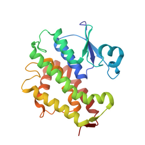
    This study presents a comprehensive analysis of the dimerization interfaces of fly GSTs through sequence alignment. Our investigation revealed GSTE1 as a particularly intriguing target, providing valuable insights into the variations within Delta and Epsilon GST interfaces. The X-ray structure of GSTE1 was determined, unveiling remarkable thermal stability and a distinctive dimerization interface. Utilizing circular dichroism, we assessed the thermal stability of GSTE1 and other Drosophila GSTs with resolved X-ray structures. The subsequent examination of GST dimer stability correlated with the dimerization interface supported by findings from X-ray structural analysis and thermal stability measurements. Our discussion extends to the broader context of GST dimer interfaces, offering a generalized perspective on their stability. This research enhances our understanding of the structural and thermodynamic aspects of GST dimerization, contributing valuable insights to the field.


  • Organizational Affiliation

    Flavour Perception: Molecular Mechanisms (Flavours), INRAE, CNRS, Université de Bourgogne, 21000 Dijon, France.


Macromolecules
Find similar proteins by:  (by identity cutoff)  |  3D Structure
Entity ID: 1
MoleculeChains Sequence LengthOrganismDetailsImage
GH14654p
A, B
224Drosophila melanogaster American nodavirus (ANV) SW-2009aMutation(s): 0 
Gene Names: 
EC: 2.5.1.18
UniProt
Find proteins for Q7KK90 (Drosophila melanogaster)
Explore Q7KK90 
Go to UniProtKB:  Q7KK90
Entity Groups  
Sequence Clusters30% Identity50% Identity70% Identity90% Identity95% Identity100% Identity
UniProt GroupQ7KK90
Sequence Annotations
Expand
  • Reference Sequence
Experimental Data & Validation

Experimental Data

  • Method: X-RAY DIFFRACTION
  • Resolution: 1.80 Å
  • R-Value Free: 0.177 
  • R-Value Work: 0.154 
  • Space Group: P 21 21 21
Unit Cell:
Length ( Å )Angle ( ˚ )
a = 81.038α = 90
b = 82.919β = 90
c = 95.543γ = 90
Software Package:
Software NamePurpose
REFMACrefinement
XDSdata reduction
Aimlessdata scaling
BALBESphasing

Structure Validation

View Full Validation Report


Ligand Structure Quality Assessment 


Entry History & Funding Information

Deposition Data


Funding OrganizationLocationGrant Number
Other governmentUniversite de Lorraine
Centre National de la Recherche Scientifique (CNRS)France--

Revision History  (Full details and data files)

  • Version 1.0: 2024-09-04
    Type: Initial release