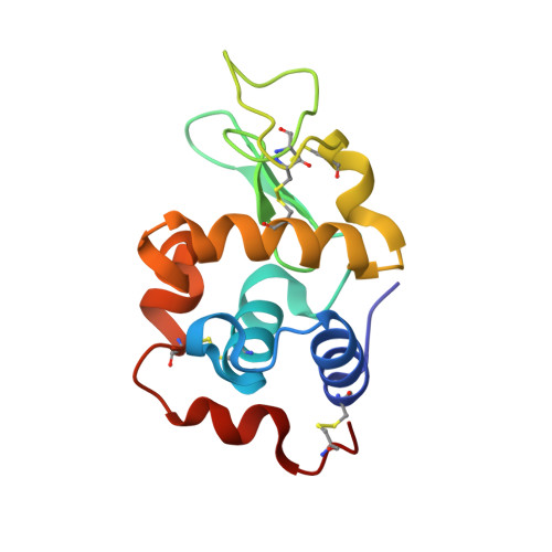
9EX1

X-ray structure of a polyoxidovanadate/lysozyme adduct obtained when the protein is treated with [VIVO(acac)2] in 1.1 M NaCl, 0.1 M sodium acetate at pH 4.0 (Structure B)


Experimental Data Snapshot

  • Method: X-RAY DIFFRACTION
  • Resolution: 1.78 Å
  • R-Value Free: 0.300 
  • R-Value Work: 0.248 

Starting Model: experimental
View more details

wwPDB Validation   3D Report Full Report


This is version 1.3 of the entry. See complete history


Literature

Non-Covalent and Covalent Binding of New Mixed-Valence Cage-like Polyoxidovanadate Clusters to Lysozyme.

Tito, G.Ferraro, G.Pisanu, F.Garribba, E.Merlino, A.

(2024) Angew Chem Int Ed Engl 63: e202406669-e202406669

  • DOI: https://doi.org/10.1002/anie.202406669
  • Primary Citation of Related Structures:  
    9EX0, 9EX1, 9EX2

  • PubMed Abstract: 

    The high-resolution X-ray structures of the model protein lysozyme in the presence of the potential drug [VIVO(acetylacetonato)2] from crystals grown in 1.1 M NaCl, 0.1 M sodium acetate at pH 4.0 reveal the binding to the protein of different and unexpected mixed-valence cage-like polyoxidovanadates (POVs): [V15O36(OH2)]5-, which non-covalently interacts with the lysozyme surface, [V15O33(OH2)]+ and [V20O51(OH2)]n- (this latter based on an unusual {V18O43} cage) which covalently bind the protein. EPR spectroscopy confirms the partial oxidation of VIV to VV and the formation of mixed-valence species. The results indicate that the interaction with proteins can stabilize the structure of unexpected - both for dimension and architecture - POVs, not observed in aqueous solution.


  • Organizational Affiliation

    University of Naples Federico II, Chemical Sciences, ITALY.


Macromolecules
Find similar proteins by:  (by identity cutoff)  |  3D Structure
Entity ID: 1
MoleculeChains Sequence LengthOrganismDetailsImage
Lysozyme C129Gallus gallusMutation(s): 0 
EC: 3.2.1.17
UniProt
Find proteins for P00698 (Gallus gallus)
Explore P00698 
Go to UniProtKB:  P00698
Entity Groups  
Sequence Clusters30% Identity50% Identity70% Identity90% Identity95% Identity100% Identity
UniProt GroupP00698
Sequence Annotations
Expand
  • Reference Sequence
Experimental Data & Validation

Experimental Data

  • Method: X-RAY DIFFRACTION
  • Resolution: 1.78 Å
  • R-Value Free: 0.300 
  • R-Value Work: 0.248 
  • Space Group: P 43 21 2
Unit Cell:
Length ( Å )Angle ( ˚ )
a = 77.8α = 90
b = 77.8β = 90
c = 35.35γ = 90
Software Package:
Software NamePurpose
REFMACrefinement
autoPROCdata reduction
autoPROCdata scaling
PHASERphasing

Structure Validation

View Full Validation Report


Entry History & Funding Information

Deposition Data


Funding OrganizationLocationGrant Number
Ministero dell Universita e della RicercaItaly2022JMFC3X

Revision History  (Full details and data files)

  • Version 1.0: 2024-06-26
    Type: Initial release
  • Version 1.1: 2024-07-03
    Changes: Structure summary
  • Version 1.2: 2024-07-31
    Changes: Database references
  • Version 1.3: 2024-10-23
    Changes: Structure summary