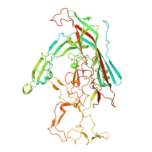
Structural Characterization of Human Bufavirus 1: Receptor Binding and Endosomal pH-Induced Changes.
Gulkis, M., Luo, M., Chipman, P., Mietzsch, M., Soderlund-Venermo, M., Bennett, A., McKenna, R.(2024) Viruses 16
- PubMed: 39205232 Search on PubMedSearch on PubMed Central
- DOI: https://doi.org/10.3390/v16081258
- Primary Citation Related Structures:
9CUZ, 9CV0, 9CV9, 9CWS - PubMed Abstract:
Bufaviruses (BuV) are members of the Parvoviridae of the Protoparvovirus genus. They are non-enveloped, T = 1 icosahedral ssDNA viruses isolated from patients exhibiting acute diarrhea. The lack of treatment options and a limited understanding of their disease mechanisms require studying these viruses on a molecular and structural level. In the present study, we utilize glycan arrays and cell binding assays to demonstrate that BuV1 capsid binds terminal sialic acid (SIA) glycans. Furthermore, using cryo-electron microscopy (cryo-EM), SIA is shown to bind on the 2/5-fold wall of the capsid surface. Interestingly, the capsid residues stabilizing SIA binding are conserved in all human BuVs identified to date. Additionally, biophysical assays illustrate BuV1 capsid stabilization during endo-lysosomal (pH 7.4-pH 4) trafficking and capsid destabilization at pH 3 and less, which correspond to the pH of the stomach. Hence, we determined the cryo-EM structures of BuV1 capsids at pH 7.4, 4.0, and 2.6 to 2.8 Å, 3.2 Å, and 2.7 Å, respectively. These structures reveal capsid structural rearrangements during endo-lysosomal escape and provide a potential mechanism for this process. The structural insights gained from this study will add to the general knowledge of human pathogenic parvoviruses. Furthermore, the identification of the conserved SIA receptor binding site among BuVs provides a possible targetable surface-accessible pocket for the design of small molecules to be developed as anti-virals for these viruses.
- Department of Biochemistry and Molecular Biology, University of Florida, Gainesville, FL 32611, USA.
Organizational Affiliation: