9CPH | pdb_00009cph

Structural basis of BAK sequestration by MCL-1 and consequences for apoptosis initiation


Experimental Data Snapshot

  • Method: ELECTRON MICROSCOPY
  • Resolution: 3.34 Å
  • Aggregation State: PARTICLE 
  • Reconstruction Method: SINGLE PARTICLE 

wwPDB Validation   3D Report Full Report


This is version 1.0 of the entry. See complete history


Literature

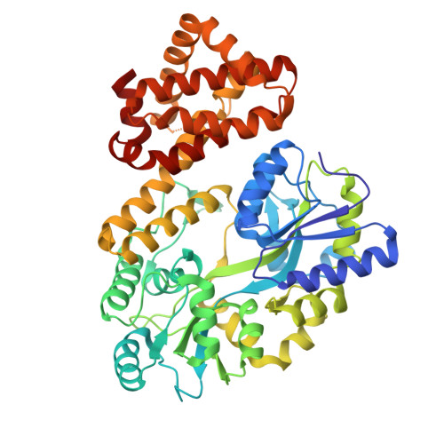
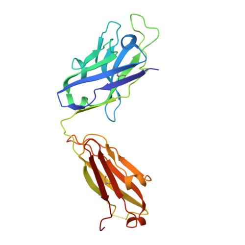
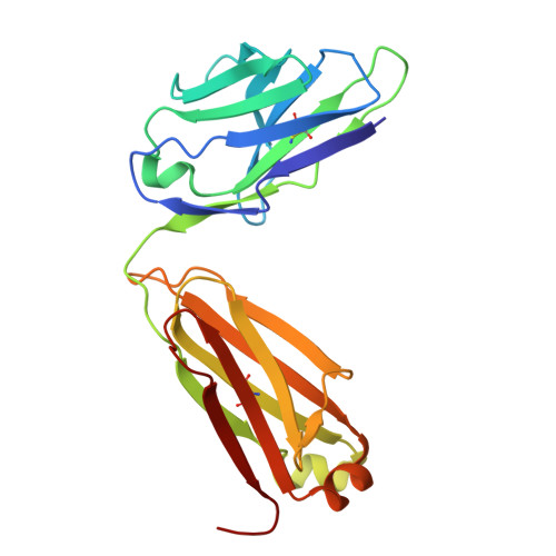
Structural basis of BAK sequestration by MCL-1 in apoptosis.

Srivastava, S.Sekar, G.Ojoawo, A.Aggarwal, A.Ferreira, E.Uchikawa, E.Yang, M.Grace, C.R.Dey, R.Lin, Y.L.Guibao, C.D.Jayaraman, S.Mukherjee, S.Kossiakoff, A.A.Dong, B.Myasnikov, A.Moldoveanu, T.

(2025) Mol Cell 85: 1606-1623.e10

  • DOI: https://doi.org/10.1016/j.molcel.2025.03.013
  • Primary Citation of Related Structures:  
    9CPE, 9CPF, 9CPH, 9CPN

  • PubMed Abstract: 

    Apoptosis controls cell fate, ensuring tissue homeostasis and promoting disease when dysregulated. The rate-limiting step in apoptosis is mitochondrial poration by the effector B cell lymphoma 2 (BCL-2) family proteins BAK and BAX, which are activated by initiator BCL-2 homology 3 (BH3)-only proteins (e.g., BIM) and inhibited by guardian BCL-2 family proteins (e.g., MCL-1). We integrated structural, biochemical, and pharmacological approaches to characterize the human prosurvival MCL-1:BAK complex assembled from their BCL-2 globular core domains. We reveal a canonical interaction with BAK BH3 bound to the hydrophobic groove of MCL-1 and disordered and highly dynamic BAK regions outside the complex interface. We predict similar conformations of activated effectors in complex with other guardians or effectors. The MCL-1:BAK complex is a major cancer drug target. We show that MCL-1 inhibitors are inefficient in neutralizing the MCL-1:BAK complex, requiring high doses to initiate apoptosis. Our study underscores the need to design superior clinical candidate MCL-1 inhibitors.


  • Organizational Affiliation
    • Biochemistry and Molecular Biology, University of Arkansas for Medical Sciences, Little Rock, AR 72205, USA.

Macromolecules
Find similar proteins by:  (by identity cutoff)  |  3D Structure
Entity ID: 1
MoleculeChains Sequence LengthOrganismDetailsImage
Induced myeloid leukemia cell differentiation protein Mcl-1515Homo sapiensMutation(s): 0 
Entity Groups  
Sequence Clusters30% Identity50% Identity70% Identity90% Identity95% Identity100% Identity
Sequence Annotations
Expand
  • Reference Sequence

Find similar proteins by:  Sequence   |   3D Structure  

Entity ID: 2
MoleculeChains Sequence LengthOrganismDetailsImage
Bcl-2 homologous antagonist/killer21Homo sapiensMutation(s): 0 
Gene Names: BAK1BAKBCL2L7CDN1
UniProt & NIH Common Fund Data Resources
Find proteins for Q16611 (Homo sapiens)
Explore Q16611 
Go to UniProtKB:  Q16611
PHAROS:  Q16611
GTEx:  ENSG00000030110 
Entity Groups  
Sequence Clusters30% Identity50% Identity70% Identity90% Identity95% Identity100% Identity
UniProt GroupQ16611
Sequence Annotations
Expand
  • Reference Sequence
Find similar proteins by:  (by identity cutoff)  |  3D Structure
Entity ID: 3
MoleculeChains Sequence LengthOrganismDetailsImage
Synthetic antibody, Fab fragment, Heavy ChainC [auth H]230Homo sapiensMutation(s): 0 
Entity Groups  
Sequence Clusters30% Identity50% Identity70% Identity90% Identity95% Identity100% Identity
Sequence Annotations
Expand
  • Reference Sequence
Find similar proteins by:  (by identity cutoff)  |  3D Structure
Entity ID: 4
MoleculeChains Sequence LengthOrganismDetailsImage
Synthetic antibody, Fab fragment, Light ChainD [auth L]214Homo sapiensMutation(s): 0 
Entity Groups  
Sequence Clusters30% Identity50% Identity70% Identity90% Identity95% Identity100% Identity
Sequence Annotations
Expand
  • Reference Sequence
Oligosaccharides

Help

Entity ID: 5
MoleculeChains Length2D Diagram Glycosylation3D Interactions
alpha-D-glucopyranose-(1-4)-alpha-D-glucopyranoseE [auth C]2N/A
Glycosylation Resources
GlyTouCan:  G07411ON
GlyCosmos:  G07411ON
Biologically Interesting Molecules (External Reference) 1 Unique
Experimental Data & Validation

Experimental Data

  • Method: ELECTRON MICROSCOPY
  • Resolution: 3.34 Å
  • Aggregation State: PARTICLE 
  • Reconstruction Method: SINGLE PARTICLE 
EM Software:
TaskSoftware PackageVersion
MODEL REFINEMENTPHENIX1.20.1_4487:

Structure Validation

View Full Validation Report


Entry History & Funding Information

Deposition Data


Funding OrganizationLocationGrant Number
National Institutes of Health/National Institute of General Medical Sciences (NIH/NIGMS)United StatesR01GM129470

Revision History  (Full details and data files)

  • Version 1.0: 2025-06-04
    Type: Initial release